Danh mục: BSNT NỘI

  • Điều trị xơ gan mất bù và ngăn ngừa tiến triển

    ĐẠI CƯƠNG

    • Điều trị lý tưởng ở bệnh nhân xơ gan mất bù là ngăn ngừa xơ gan tiến triển, không phải là điều trị biến chứng
    • Điều trị tối ưu xơ gan mất bù chủ yếu nhằm vào những thay đổi bệnh lý trong gan với mục đích khôi phục lại tính toàn vẹn cấu trúc gan bằng cách
      • ức chế viêm
      • đẩy lùi xơ hóa
      • cân đối tuần hoàn cửa và động mạch
      • bình thường hóa số lượng và chức năng tế bào

    Tế bào bị tổn thương Mô hình phân tử liên quan tổn thương

    Những cơ chế

    khác

    Tăng áp cửa

    Kích hoạt

    thụ thể nhận dạng bẩm sinh

    Sự chuyển chỗ vi khuẩn

    Tổn thương gan

    Phóng thích các phân tử tiền viêm

    ++

    Giãn tiểu động mạch tạng, rối loạn chức năng tim mạch

    năng thận

    Rối loạn chức năng thượng thận

    Sinh bệnh của xơ gan mất bù

    Bernardi M. J Hepatol 2015;63:1272–1284

    Xơ gan

    Bệnh não gan Rối loạn chức

    Hội chứng gan thận

    Tổn thương thận cấp

    Báng bụng

    HCGT

    Vàng da

    Suy gan

    Bệnh não gan

    Xơ gan mất bù

    Tăng áp cửa

    Xơ gan

    còn bù

    XHTH

    VPMNKNP

    HCC

    HCC

    1 điểm 2 điểm 3 điểm
    Bệnh não gan Báng bụng

    Bilirubin máu

    Albumin máu TQ kéo dài

    không không

    < 2 mg/dl

    > 3,5 g/dl

    < 4’’

    độ 1–2 ít 2–3

    2,8–3,5

    4–6”

    độ 3–4

    trung bình, nhiều

    > 3

    < 2,8

    > 6”

    Hoặc INR < 1,7 1,7–2,2 > 2,2
    XG ứ mật Bili < 4 4–10 > 10

    KHẢ NĂNG SỐNG CÒN NGUY CƠ PHẪU THUẬT THEO CHILD-PUGH

    Child-Pugh A B C
    Điểm CTP 5–6 7–9 10–15
    Tuổi thọ (năm) 15–20 4–14 1–3
    Tử vong chu phẫu (%) 10 30 80

    Schuppan D, Afdhal NH. Liver cirrhosis 2008

    1. EASL clinical practice guidelines on the management of ascites, spontaneous bacterial peritonitis, and hepatorenal syndrome in cirrhosis. J Hepatol 2010;53:397–417
    2. Lach J, et al. Prognostic value of arterial pressure, endogenous vasoactive systems, and renal function in cirrhotic patients admitted to the hospital for the treatment of ascites. Gastroenterology 1988;94:482–487.
    3. Schuppan D, Afdhal NH. Liver cirrhosis 2008
    4. Caregaro L, et al. Limitations of serum creatinine level and creatinine clearance as filtration markers in cirrhosis. Arch Intern Med 1994;154:201–205. 9
    5. Bernardi M, et al. The MELD score in patients awaiting liver transplant: strengths and weaknesses. J Hepatol 2011;54:1297–1306.
    • Xơ gan có báng bụng tiên lượng xấu, tỷ lệ tử vong 1 và 2 năm ♯ 40 và 50%, theo thứ tự 1
    • Hạ natri máu, huyết áp động mạch thấp, độ lọc cầu thận và bài tiết Na niệu thấp: yếu tố tiên lượng tử vong độc lập ở bệnh nhân xơ gan báng bụng 2
    • Điểm Child-Pugh 3
    • Mô hình bệnh gan giai đoạn cuối (MELD) 4
    • Điểm MELD-Na và MELD-báng bụng 5
    • Tùy thuộc biến chứng
    • Điều trị chung xơ gan mất bù:
      1. Ngăn chặn các yếu tố căn nguyên
      2. Tác động lên các yếu tố đích của bệnh sinh mất bù và tiến triển của xơ gan
    • Loại bỏ các yếu tố căn nguyên gây tổn thương gan
      • Nền tảng quan trọng trong điều trị xơ gan
      • Hiệu quả trong việc ngăn ngừa mất bù
      • Cải thiện kết cục ở bệnh nhân xơ gan còn bù

    10

    ĐIỀU TRỊ XƠ GAN MẤT BÙ

    EASL Clinical Practice Guidelines: Management of alcohol-related liver disease.

    Journal of Hepatology 2018 vol. 69 j 154–181

    • Các bước tiếp cận giảm tiến triển của xơ gan
      • Cải thiện trục gan-ruột nhằm vào bất thường vi sinh vật và sự chuyển chỗ vi khuẩn bằng kháng sinh (rifaximin)
      • Cải thiện chức năng tuần hoàn hệ thống bị rối loạn (albumin)
      • Giảm tình trạng viêm (statin)
      • Giảm tình trạng tăng áp cửa (thuốc chẹn beta)
    • Điều trị triệu chứng và biến chứng

    11
    ĐIỀU TRỊ BÁNG BỤNG DO XƠ GAN

    12

    1. Moore KP, Wong F, Gines P, Bernardi M, Ochs A, Salerno F, et al. The management of ascites in cirrhosis: report on the consensus conference of the International Ascites Club. Hepatology 2003;38:258–266.

    2. Arroyo V, Gines P, Gerbes AL, Dudley FJ, Gentilini P, Laffi G, et al. Definition and diagnostic criteria of refractory ascites and hepato1r3enal

    syndrome in cirrhosis. International Ascites Club. Hepatology 1996;23:164–176.

    • Xơ gan bị báng bụng độ 2 hoặc 3 sống còn giảm

     cân nhắc ghép gan khi xơ gan có báng bụng

    • Báng bụng không biến chứng khi không bị

    nhiễm trùng, kháng trị hoặc hội chứng gan thận

    • Phân độ báng bụng
      • Độ 1 – ít: phát hiện nhờ siêu âm
      • Độ 2 – trung bình: bụng chướng vừa phải
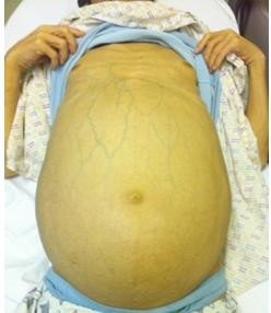
      • Độ 3 – lượng lớn: bụng chướng căng rõ

    Chẩn đoán báng bụng

    • Chọc dịch báng chẩn đoán tất cả bệnh nhân báng bụng độ 2 – 3 hoặc nhập viện vì báng bụng xấu hơn hoặc vì bất kỳ biến chứng nào của xơ gan
    • Đo protein dịch báng để xác định nguy cơ VPMNKNP
    • SAAG khi không rõ nguyên nhân báng bụng và/hoặc nghi ngờ nguyên nhân khác gây báng bụng
    • Đếm tế bào để phân biệt báng bụng do bệnh ác tính

    14

    • Báng bụng độ 2 không biến chứng
      • Nghỉ ngơi/giường có lợi trong điều trị báng bụng?
      • Hạn chế natri
        • Hạn chế natri vừa phải (80–120 mmol/ngày, tương ứng 4,6-6,9 g muối), tương đương với chế độ ăn không có muối và tránh các bữa ăn được chuẩn bị trước
        • Tránh chế độ ăn có lượng natri rất thấp (<40 mmol/ngày) vì dễ bị các biến chứng do lợi tiểu và không tốt cho dinh dưỡng
        • Hướng dẫn dinh dưỡng đầy đủ
    • Báng bụng độ 2
      • Thuốc lợi tiểu
        • Báng bụng độ 2, lần đầu:

    sử dụng 1 thuốc kháng mineralocorticoid

    bắt đầu 100 mg/ngày, tăng dần 100 mg/ 72 giờ tối đa 400 mg/ngày nếu không đáp ứng

    Nếu không đáp ứng ( cân <2 kg/tuần) hoặc  kali máu, thêm furosemide 40 mg/ngày tăng dần đến tối đa 160 mg/ngày

        • Báng bụng kéo dài/ tái phát: kháng mineralo- corticoid và furosemide, tăng liều tùy đáp ứng
        • Đáp ứng furosemide kém, sử dụng Torasemide
    • Báng bụng độ 2
      • Thuốc lợi tiểu
        • Giảm cân tối đa 0,5 kg/ngày nếu không phù và 1 kg/ngày nếu có phù
        • Khi kiểm soát được báng bụng, nên giảm liều thuốc lợi tiểu đến mức thấp nhất có hiệu quả
        • Trong những tuần đầu điều trị, cần theo dõi lâm sàng và sinh hóa máu thường xuyên, nhất là khi báng bụng lần đầu
    • Báng bụng độ 2
      • Ngừng thuốc lợi tiểu
        • Ngừng thuốc lợi tiểu nếu: natri máu <125 mmol/L, TTTC, bệnh não gan xấu hơn / kéo dài, chuột rút
        • Ngừng furosemide, nếu K+ máu <3 mmol/L
        • Ngừng kháng mineralocorticoid, nếu K+ >6 mmol/L
      • Truyền albumin hoặc baclofen (10 mg/ngày, mỗi tuần tăng 10 mg/ngày, đến 30 mg/ngày) đối với những bệnh nhân bị chuột rút

    18

    • Báng bụng độ 3 (lượng lớn)
      • Chọc tháo dịch báng lượng lớn: xử trí hàng đầu đối với báng bụng lượng lớn, nên chọc tháo toàn bộ trong một lần duy nhất
      • Chọc tháo dịch báng <5 lít, nguy cơ rối loạn tuần hoàn thấp. Chọc tháo dịch báng >5 L, nên tăng thể tích huyết tương bằng truyền albumin (8 g/L dịch báng), hiệu quả hơn so với các dịch tăng thể tích huyết tương khác
      • Sau khi chọc tháo dịch báng, nên sử dụng lợi tiểu liều tối thiểu để ngăn ngừa báng bụng tái phát
    • Chống chỉ định chọc tháo dịch báng
      • Bệnh nhân không hợp tác
      • Nhiễm trùng da vùng bụng tại vị trí chọc tháo
      • Có thai
      • Bệnh đông máu nặng (tăng tiêu sợi huyết hoặc đông máu nội mạch lan tỏa)
      • Chướng ruột nặng
    • Thuốc sử dụng đồng thời trong xơ gan báng bụng
      • Không nên sử dụng NSAID vì có nguy cơ cao giữ natri, hạ natri máu và tổn thương thận cấp (TTTC)
      • Không nên sử dụng các thuốc ức chế men chuyển angiotensin, đối kháng angiotensin II hoặc chẹn thụ thể α1-adrenergic vì tăng nguy cơ suy thận
      • Không nên sử dụng kháng sinh aminoglycoside, vì tăng nguy cơ TTTC. Ngoại trừ trường hợp nhiễm khuẩn nặng không thể điều trị bằng các thuốc khác
      • XGBB và chức năng thận bảo tồn, thuốc cản quang không liên quan đến tăng nguy cơ suy thận
    Định nghĩa
    Báng bụng kháng trị thuốc lợi tiểu

    Diuretic-resistant ascites

    Báng bụng không cải thiện/ tái phát sớm vì thiếu đáp ứng với hạn chế natri và điều trị lợi tiểu
    Báng bụng khó chữa với thuốc lợi tiểu

    Diuretic-intractable ascites

    Báng bụng không cải thiện/ tái phát sớm vì có các biến chứng do thuốc lợi tiểu (ngăn cản việc sử dụng liều thuốc lợi tiểu hiệu quả)
    Tiêu chuẩn chẩn đoán
    Thời gian điều trị Bệnh nhân phải điều trị lợi tiểu liều cao (spironolactone 400 mg/ngày và furosemide 160 mg/ngày) ít nhất một tuần và chế độ ăn muối <90 mmol/ngày
    Thiếu đáp ứng Giảm cân <0,8 kg/ 4 ngày và

    lượng natri niệu < lượng natri ăn vào

    Báng bụng tái phát sớm Báng bụng độ 2 hoặc 3 tái phát trong vòng 4 tuần kể từ khi bắt đầu điều trị
    Tiêu chuẩn chẩn đoán Báng bụng kháng trị
    Biến chứng do thuốc lợi tiểu
    • Bệnh não gan do thuốc lợi tiểu, không có bất kỳ yếu tố thúc đẩy nào khác
    • Suy thận do thuốc lợi tiểu: creatinine máu tăng >100% đến giá trị >2 mg/dl ở bệnh nhân báng bụng đáp ứng với điều trị
    • Hạ natri máu do thuốc lợi tiểu: giảm natri máu nhiều hơn 10 mmol/L đến giá trị natri máu <125 mmol/L
    • Tăng hoặc giảm kali máu do thuốc lợi tiểu: kali máu >6 mmol/L hoặc <3 mmol/L
    • Chuột rút

    24

    • Chọc tháo dịch báng lượng lớn lặp lại kèm albumin: điều trị hàng đầu đối với báng bụng kháng trị
    • Nên ngừng thuốc lợi tiểu ở bệnh nhân báng bụng kháng trị bài tiết natri niệu ≤30 mmol /ngày
    • Thận trọng khi sử dụng NSBB trong trường hợp báng bụng nặng hoặc kháng trị. Tránh sử dụng NSBB liều cao (propranolol >80 mg/ngày). Không khuyến cáo sử dụng carvedilol

    NSBB: NonSelective Beta Blockers

    • Bệnh nhân bị báng bụng kháng trị hoặc tái phát, hoặc chọc tháo không hiệu quả nên xem xét việc đặt shunt cửa chủ trong gan (TIPS)
    • Đặt TIPS giúp cải thiện sống còn ở bệnh nhân báng bụng tái phát và cải thiện việc kiểm soát báng bụng ở bệnh nhân báng bụng kháng trị
    • Sử dụng stent đường kính nhỏ, có phủ PTFE để giảm nguy cơ bệnh não gan và rối loạn chức năng do TIPS
    • Thuốc lợi tiểu và hạn chế natri nên được tiếp tục sau đặt TIPS

    TIPS: Transjugular Intrahepatic Portosystemic Shunt

    • TIPS không được khuyến cáo trong trường hợp
      • Bilirubin huyết thanh >3 mg/dl
      • Số lượng tiểu cầu <75 x 109/L,
      • Bệnh não gan độ ≥2 hoặc bệnh não gan mạn
      • Nhiễm trùng đang hoạt động
      • Suy thận tiến triển
      • Tăng áp phổi hoặc rối loạn chức năng tâm trương hoặc tâm thu nặng

    ĐIỀU TRỊ

    VIÊM PHÚC MẠC NHIỄM KHUẨN NGUYÊN PHÁT

    28
    Nguy cơ nhiễm khuẩn trong xơ gan do nhiều yếu tố

    • Rối loạn chức năng gan
    • Thông nối cửa chủ
    • Mất cân bằng vi sinh vật ở ruột
    • Tăng chuyển chỗ vi khuẩn
    • Rối loạn chức năng miễn dịch do xơ gan
    • Những yếu tố di truyền
    • Chọc dịch báng chẩn đoán VPMNKNP ở BN XGBB
      • Lúc nhập viện để loại trừ VPMNKNP
      • Bị xuất huyết tiêu hóa, sốc, sốt, có những dấu hiệu nhiễm khuẩn toàn thân, triệu chứng tiêu hóa
      • Tình trạng gan và thận xấu hơn, bệnh não gan
    • Nghi ngờ VPMNK thứ phát nếu
      • cấy dịch báng nhiều loại vi khuẩn
      • số lượng bạch cầu trung tính rất cao
      • nồng độ protein dịch báng cao
      • hoặc đáp ứng kém với điều trị
    • Đếm số lượng bạch cầu trung tính dịch báng >250

    /mm3 là cần thiết để chẩn đoán VPMNKNP

    • Cấy dịch báng tại giường không phải là điều kiện tiên quyết để chẩn đoán VPMNKNP, tuy nhiên giúp hướng dẫn điều trị kháng sinh
    • Cấy máu nên được thực hiện ở tất cả bệnh nhân nghi ngờ VPMNKNP, trước khi điều trị kháng sinh
    • Bệnh nhân bị du khuẩn báng (bạch cầu trung tính

    <250/mm3, cấy (+)) có dấu hiệu nhiễm trùng hoặc đáp ứng viêm toàn thân nên điều trị kháng sinh.

    Nếu không, phải chọc dịch lần hai. Cấy lần hai (+), bất kể số lượng bạch cầu, nên điều trị kháng sinh

    Jalan R, et al. Bacterial infections in cirrhosis: a position statement based on

    the EASL Special Conference. J Hepatol 2013;60:1310–1324

    32

    Phụ thuộc vùng: giống nhiễm trùng bệnh viện nếu xuất độ vi khuẩn kháng đa thuốc cao hoặc nhiễm trùng huyết

    VPMNKNP

    do chăm sóc y tế

    Carbapenem đơn thuần hoặc phối hợp daptomycin, vancomycin hoặc linezold nếu xuất độ vi khuẩn gram + kháng đa thuốc hoặc nhiễm trùng huyết

    VPMNKNP

    bệnh viện

    VPMNKNP: viêm phúc mạc nhiễm khuẩn nguyên phát MDRO: vi khuẩn kháng đa thuốc

    Cephalosporin thế hệ 3 hoặc piperacillin- tazobactam

    VPMNKNP

    mắc phải ở cộng đồng

    VPMNKNP

    • Kháng sinh theo kinh nghiệm bắt đầu ngay lập tức.
    • Kháng sinh theo hướng dẫn bởi đặc tính đề kháng của vi khuẩn
    • VPMNKNP cộng đồng, tùy vùng:

    Tỷ lệ kháng khuẩn thấp: cephalosporin 3

    Tỷ lệ kháng khuẩn cao: piperacillin/tazobactam hoặc carbapenem

    • VPMNKNP do chăm sóc y tế & bệnh viện, tùy vùng: Tỷ lệ kháng đa thuốc thấp: piperacillin/tazobactam Tỷ lệ cao vi khuẩn sinh  lactamase: carbapenem Tỷ lệ vi khuẩn Gr (+) đa kháng thuốc cao, phối hợp carbapenem với glycopeptide hoặc daptomycin

    hoặc linezolid

    33

    Jalan R, et al. Bacterial infections in cirrhosis: a position statement based on

    the EASL Special Conference. J Hepatol 2013;60:1310–1324

    • Kiểm tra hiệu quả của điều trị kháng sinh: xét nghiệm dịch báng lần hai tại thời điểm 48 giờ sau điều trị. Nghi ngờ thất bại điều trị kháng sinh nếu triệu chứng và dấu hiệu lâm sàng xấu hơn và/hoặc số lượng bạch cầu  hoặc không giảm rõ (ít nhất 25%) sau 48 giờ
    • Thời gian điều trị ít nhất 5-7 ngày
    • Mủ màng phổi nguyên phát được điều trị tương tự viêm phúc mạc nhiễm khuẩn nguyên phát
    • Albumin (1,5 g/kg lúc chẩn đoán và 1 g/kg vào ngày thứ 3) được khuyến cáo ở bệnh nhân bị VPMNKNP

    34

    Dự phòng tiên phát: bệnh nhân có protein dịch báng <15 g/L, không có tiền căn bị VPMNKNP

    • Dự phòng tiên phát với norfloxacin (400 mg/ngày) ở bệnh nhân
      • Child-Pugh ≥9 và bilirubin huyết thanh ≥3 mg/dl, chức năng thận giảm hoặc
      • Hạ natri máu
    • Ngừng dự phòng Norfloxacin ở bệnh nhân có cải thiện lâm sàng kéo dài và hết báng bụng

    Bệnh nhân bị VPMNKNP: dự phòng tái phát

    • Norfloxacin (400 mg/ngày, uống) cho bệnh nhân hồi phục sau VPMNKNP
    • Rifaximin không được khuyến cáo thay thế norfloxacin để dự phòng VPMNKNP tái phát
      • Sau đợt điều trị VPMNKNP
        • không xác định
        • hết báng bụng
      • Bệnh nhân nguy cơ cao
        • trong thời gian nằm viện
      • Đang bị xuất huyết tiêu hóa
        • 7 ngày
    • Sau đợt điều trị VPMNKNP
      • Norfloxacin 400 mg/ngày
      • Ciprofloxacin 500-1.000 mg/ngày
      • Trimethoprim-Sulfamethoxazole 960mg/ngày
    • Đang bị XHTH
      • Norfloxacin 400 mg X 2 lần/ngày X 7 ngày
      • Ceftriaxone 1 g tiêm mạch/ngày X 7 ngày

    EASL Clinical Practice Guidelines for the management of patients with decompensated cirrhos3is9.

    Journal of Hepatology 2018 vol. xxx

    • PPI có thể làm tăng nguy cơ bị VPMNKNP, nên giới hạn sử dụng đối với người có chỉ định rõ
    • NSBB có thể gây bất lợi cho bệnh gan giai đoạn cuối bị rối loạn huyết động, cần theo dõi sát và điều chỉnh liều hoặc ngừng thuốc nếu có chống chỉ định
    • Probiotic: không có bằng chứng có lợi
    • Bệnh nhân hồi phục sau VPMNKNP có sống còn kém, cần cân nhắc để ghép gan
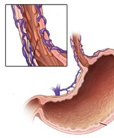
    A picture containing indoor  Description generated with high confidence

    PHÒNG NGỪA XUẤT HUYẾT TIÊU HÓA

    DO VỠ TĨNH MẠCH GIÃN

    40

    XUẤT HUYẾT TIÊU HÓA

    1. North Italian Endoscopic Club for the Study and Treatment of Esophageal Varices. Prediction of the first variceal hemorrhage in patients with cirrhosis of the liver and esophageal varices. A prospective multicenter study. N Engl J Med 1988;319:983–989.
    2. D’Amico G, et al. Competing risks and prognostic stages of cirrhosis: a 25-year inception cohort study of 494 patients. Aliment Pharmacol Ther 2014;39:1180–1193.
    3. Augustin S, et al. Predicting early mortality after acute variceal hemorrhage based on classification and regression tree analysis. Clin Gastroenterol Hepatol 2009;7:1347– 1354. 41
    4. Bosch J, et al. Prevention of variceal rebleeding. Lancet 2003;361:952–954
    • Tỉ lệ tử vong chung mỗi đợt xuất huyết do tĩnh mạch giãn 15%-25% vào tuần thứ sáu. Nguy cơ tử vong ở những người xuất huyết kèm biểu hiện mất bù khác (>80% ở thời điểm 5 năm) cao hơn nhiều so với xuất huyết do tĩnh mạch giãn là biến cố mất bù đơn độc (20% ở thời điểm 5 năm) 6,7
    • Nguy cơ tử vong cao khi kèm TTTC, VPMNKNP 8
    • Không phòng ngừa, xuất huyết tái phát 60%-70%, thường trong vòng 1-2 năm sau xuất huyết 9

    Tiếp cận điều trị xuất huyết tiêu hóa cấp ở bệnh nhân xơ gan

    Xuất huyết tiêu hóa cấp + Tăng áp cửa

    Đánh giá ban đầu

    Airway Breathing Circulation

    Bồi hoàn thể tích bằng dung dịch tinh thể/keo) Truyền máu duy trì Hb 7 g/dl, mục tiêu 7-9 g/dl

    Chèn bóng/đặt stent thực quản

    nếu xuất huyết lượng lớn

    Điều trị ngay: somatostatin, terlipressin

    KS phòng ngừa: ceftriaxone, norfloxacine

    Nội soi chẩn đoán sớm (<12 giờ) Xác định xuất huyết do tĩnh mạch giãn

    Điều trị qua nội soi (thắt tĩnh mạch)

    + duy trì thuốc co mạch 3-5 ngày

    và phòng ngừa kháng sinh

    De Franchis R. Expanding consensus in portal hypertension: report of the BAVENO VI Consensus Workshop: Stratifying risk and individualizing care for portal hypertension. J Hepa4t2ol

    2015;63:743–752

    Kiểm soát (~85%) Còn xuất huyết (~15%)

    Cân nhắc TIPS sớm

    TIPS cứu vãn
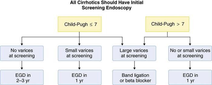
      • Nội soi
        • Thắt TMTQ
        • Chích xơ TM phình vị
      • Thuốc chẹn beta

    Nên nội soi thường qui

    cho tất cả BNXG lần đầu

    Child Pugh  7

    Child Pugh > 7

    Không giãn TM

    Giãn TM nhỏ

    Giãn TM lớn

    Không, giãn nhỏ

    NS: 2-3 năm

    NS: 1 năm

    Thắt, chẹn beta

    NS: 1 năm

    • Tĩnh mạch giãn nhẹ có dấu son hoặc Child-Pugh C điều trị NSBB (thuốc chẹn beta không chọn lọc)
    • Tĩnh mạch giãn trung bình hoặc lớn điều trị NSBB hoặc thắt tĩnh mạch. Chọn lựa tùy khả năng tại chỗ, sự ưa thích của người bệnh, chống chỉ định và tác dụng phụ. NSBB có thể được ưa chọn hơn vì ngoài tác động giảm áp cửa còn có những tác động có lợi khác
    • Báng bụng không phải là chống chỉ định sử dụng NSBB. Thận trọng trong những trường hợp báng bụng nặng hoặc kháng trị, tránh sử dụng NSBB liều cao, không sử dụng carvedilol
      • Bệnh nhân bị hạ áp tiến triển (HAmax <90 mmHg) hoặc trong tình trạng cấp: xuất huyết, nhiễm trùng, VPMNKNP hoặc TTTC, ngừng NSBB

    Sau khi hồi phục, có thể thử dùng lại. Nếu không dung nạp hoặc chống chỉ định NSBB hoặc có nguy cơ xuất huyết nên phòng ngừa xuất huyết bằng thắt tĩnh mạch giãn

      • Phối hợp NSBB và thắt tĩnh mạch giãn giảm nguy cơ chảy máu tái phát tốt hơn so với đơn trị liệu
      • Bệnh nhân không dung nạp NSBB, xem xét đặt TIPS, nếu không có chống chỉ định tuyệt đối

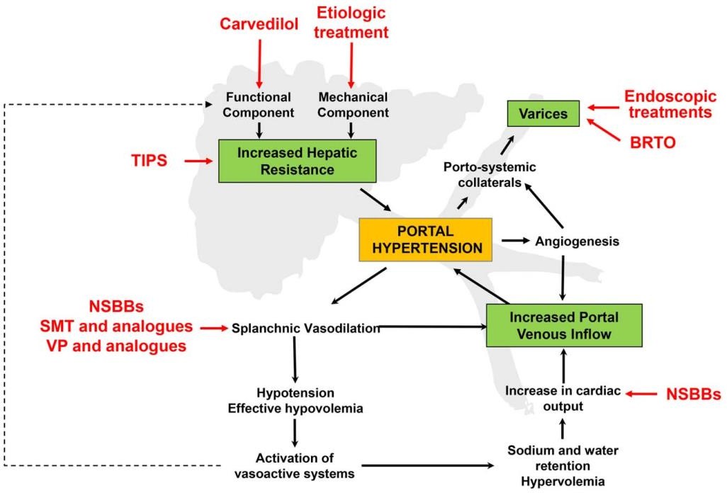
    Cơ chế bệnh sinh Tăng áp cửa và

    Vị trí hoạt động của các điều trị được khuyến cáo

    AASLD 2017
    46

    Cơ chế tác động của NSBB / XG còn bù Cơ chế tác động của NSBB / XG mất bù

    Carvedilol

    1

    1 2

    Propranolol Nadolol

    có lợi

    tiềm ẩn có hại có hại

    1 Carvedilol

    1 Propranolol

    Nadolol

    Mất khả năng ino/chomotropic

    2

    Chống lại tăng cung lượng tim

    Nhằm vào kháng trở mạch máu trong gan

    Bảo tồn ino/chomotropic

    Kháng trở trong gan

    Tim

    Giảm cung lượng tim

    Nhằm vào kháng trở mạch máu trong gan

    Dòng máu đến tạng

    Giảm giãn mạch tạng

    Giảm giãn mạch tạng

    DOI: (10.1
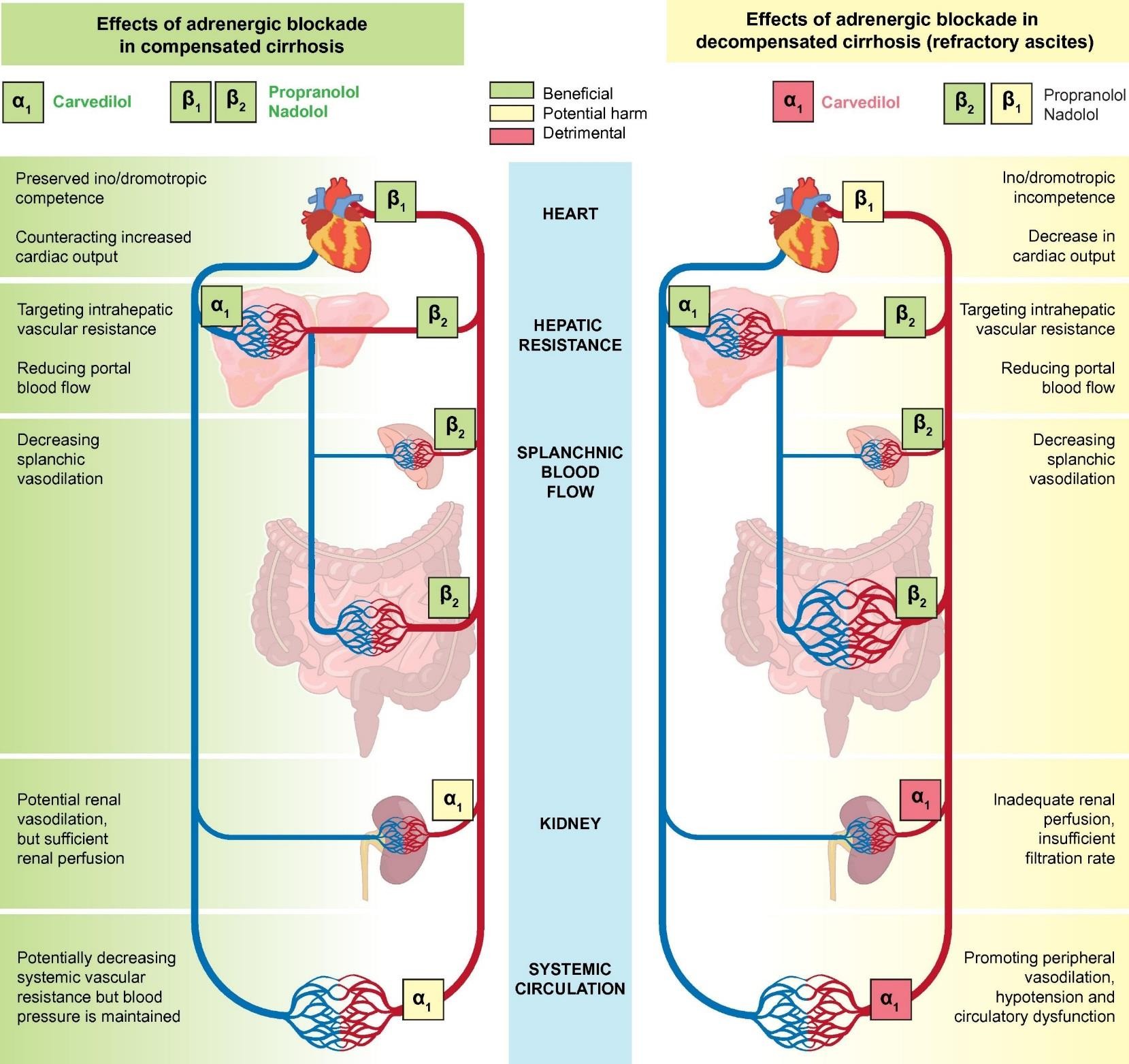
     

    Giảm dòng máu cửa

    Giảm dòng máu cửa

    Thận

    Tuần hoàn hệ thống

    Kháng trở mạch máu hệ thống giảm tiềm tàng nhưng huyết áp được duy trì.

    Tưới máu thận thiếu, tốc độ lọc không đủ

    Giãn mạch thận tiềm tàng nhưng tưới máu thận đủ

    EASL 2018

    Giãn mạch ngoại biên, hạ áp và rối loạn chức năng tuần hoàn

    ournal of Hepatology 2017 66, 859 016 2016.11.001)

    Thuốc chẹn beta không chọn lọc

    • ức chế thụ thể beta trên các mạch máu tạng

      cung lượng tim,  dòng máu tĩnh mạch cửa

      áp tĩnh mạch cửa

    •  kháng trở bàng hệ quanh hệ cửa   dòng máu bàng hệ
    •  xuất huyết khi nhịp tim  25% nhịp cơ bản
    •  nguy cơ xuất huyết tiên phát khoảng 50%
    • khoảng 1/3 bệnh nhân không dung nạp thuốc
    • không ngăn ngừa hình thành giãn tĩnh mạch

    Thuốc chẹn beta không chọn lọc (NSBB) Chống chỉ định thuốc Propranolol

      • Hen, bệnh phổi tắc nghẹn mạn (COPD)
      • Nhịp tim chậm <50 lần/phút
      • Hội chứng suy nút xoang
      • Blốc nhĩ thất độ 2 hoặc 3
      • Sốc, hạ huyết áp nặng
      • Suy tim sung huyết không kiểm soát được

    Thuốc chẹn beta không chọn lọc (NSBB) Dược động học thuốc propranolol

      • Hấp thu nhanh & hoàn toàn
      • Đạt nồng độ đỉnh 1-3 giờ sau uống
      • Tăng hoạt tính sinh học khi bị suy gan
      • Thời gian bán hủy 3-4 giờ
      • Có ái lực với mô mỡ
      • Chuyển hóa thành 4-hydroxypropranolol, có hoạt tính dược lý, thời gian bán hủy 5,2-7,2 giờ

    Thuốc chẹn beta không chọn lọc (NSBB) Tác dụng phụ

      • Buồn nôn, tiêu chảy
      • Co thắt phế quản, khó thở
      • Lạnh đầu chi, hội chứng Raynaud nặng hơn
      • Chậm nhịp tim, hạ huyết áp, suy tim
      • Mệt mỏi, chóng mặt, thị lực bất thường
      •  tập trung, ảo giác, mất ngủ, ác mộng
      • Rối loạn chuyển hóa glucose & lipid
    Thuốc Liều đầu/ngày Liều điều trị/ngày
    Propranolol * 20-40 mg x 2 160 mg có báng bụng
    320 mg báng bụng (-)
    Nadolol * 20-40 mg 80 mg có báng bụng
    160 mg báng bụng (-)
    Carvedilol * 6,25 mg 6,25 mg x 2
    • Garcia-Tsao G. Portal Hypertensive Bleeding in Cirrhosis. HEPATOLOGY, Vol. 65, No. 1,

    2017. AASLD Practice Guidelines 2017

    52

      • Carvedilol không được khuyến cáo trong
        • Phòng ngừa xuất huyết tái phát do tĩnh mạch giãn *
        • Xơ gan báng bụng nặng hoặc kháng trị **
      • Mục tiêu điều trị *
        • Nhịp tim lúc nghỉ 55-60 lần/phút
        • Huyết áp tâm thu không nên giảm <90 mmHg
    • Garcia-Tsao G. Portal Hypertensive Bleeding in Cirrhosis. HEPATOLOGY, Vol. 65, No. 1,

    2017. AASLD Practice Guidelines 2017

    ** EASL Clinical Practice Guidelines for the management of patients with decompensated

    53

    cirrhosis. Journal of Hepatology 2018 vol. xxx

    ĐIỀU TRỊ

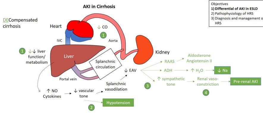
    TỔN THƯƠNG THẬN CẤP TRONG XƠ GAN

    54
    Nam, 38 t, 50 kg, 4 ngày nay: tiêu chảy, sốt, báng bụng độ 3, đau vùng rốn, nhập BVĐK tỉnh: creatinine máu 1,3 mg/dL, tiểu 100 ml/6 giờ, chẩn đoán xơ gan mất bù, sau 24 giờ chuyển BVCR. Tại BVCR: creatinine máu 1,63 mg/dL. Chẩn đoán bệnh nhân bị tổn thương thận cấp dựa tiêu chuẩn nào?

    0%

    1. Creatinine máu
    2. Lượng nước tiểu
    3. Creatinine máu và lượng nước tiểu

    0%

    0%

    Tiêu chuẩn chẩn đoán TTTC trong xơ gan

    Angeli P et al. International Club of Ascites. Gut. 2015 Apr; 64(4):531-7

    Thông số Định nghĩa

    TTTC

    Tăng Cr HT ≥26,4 μmoL/L (0,3 mg/dL) trong 48 giờ

    hoặc tăng 50% so với giá trị nền

    Thông số Định nghĩa

    Cr HT nền Creatinine huyết thanh (Cr HT ổn định ≤3 tháng)

    Nếu không có, trị số Cr HT gần nhất lần nhập viện này

    Nếu trước đó không có, sử dụng Cr HT lúc nhập viện

    TTTC: tổn thương thận cấp Cr HT: creatinine huyết thanh

    Nam, 38 t, 4 ngày nay: tiêu chảy, sốt, báng bụng độ 3, đau vùng rốn, nhập BVĐK tỉnh: creatinine máu 1,3 mg/dL, chẩn đoán xơ gan mất bù, sau 24 giờ chuyển BVCR. Tại BVCR: creatinine máu 1,63 mg/dL. Bệnh nhân bị tổn thương thận cấp giai đoạn mấy?

    0%

    1. 1
    2. 2
    3. 3

    0%

    0%

    Tiêu chuẩn chẩn đoán TTTC trong xơ gan

    Angeli P et al. International Club of Ascites. Gut. 2015 Apr; 64(4):531-7

    Thông số Định nghĩa

    Cr HT nền Creatinine huyết thanh (Cr HT ổn định ≤3 tháng)

    Nếu không có, trị số Cr HT gần nhất lần nhập viện này Nếu trước đó không có, sử dụng Cr HT lúc nhập viện

    Tổn thương thận cấp giai đoạn

    1 Tăng Cr HT ≥26,4 μmoL/L (0,3 mg/dL) hoặc tăng Cr HT ≥1,5–2 × giá trị nền

    2 Tăng Cr HT >2–3 × giá trị nền

    3 Tăng Cr HT >3 × giá trị nền hoặc

    Cr HT ≥352 μmoL/L (4 mg/dL) có ↑ cấp ≥26,4 μmoL/L

    hoặc bắt đầu điều trị thay thế thận

    Cr HT: creatinine huyết thanh

    Nam, 38 t, 4 ngày nay: tiêu chảy, sốt, báng bụng độ 3, đau vùng rốn, nhập BVĐK tỉnh: creatinine máu 1,3 mg/dL, chẩn đoán xơ gan mất bù, sau 24 giờ chuyển BVCR. Tại BVCR: creatinine máu 1,63 mg/dL, sau 48 giờ: creatinine máu 1 mg/dL. Đáp ứng điều trị của tổn thương thận cấp thuộc nhóm nào?

    0%

    1. Không đáp ứng
    2. Đáp ứng một phần
    3. Đáp ứng hoàn toàn

    0%

    0%

    Định nghĩa đáp ứng của TTTC

    Angeli P et al. International Club of Ascites. Gut. 2015 Apr; 64(4):531-7

    Đáp ứng điều trị

    Không TTTC không thoái lui

    Một phần

    Giai đoạn TTTC thoái lui với Cr HT giảm ≤0,3

    mg/dL so với giá trị nền

    Hoàn toàn Giảm Cr HT >0,3 mg/dL so với giá trị nền

    Định nghĩa

    Cr HT nền Creatinine huyết thanh (Cr HT ổn định ≤3 tháng)

    Nếu không có, trị số Cr HT gần nhất lần nhập viện này Nếu trước đó không có, sử dụng Cr HT lúc nhập viện

    60

    TTTC: tổn thương thận cấp Cr HT: creatinine huyết thanh

    Nam, 38 t, 4 ngày nay: tiêu chảy, sốt, báng bụng độ 3, khó thở khi nằm, đau vùng rốn, nhập BVĐK tỉnh: creatinine máu 1,3 mg/dL, uống Aldacton 100 mg và furosemide 40 mg, chẩn đoán xơ gan mất bù, sau 24 giờ chuyển BVCR. Tại BVCR: creatinine máu 1,63 mg/dL, K+ 3 mEq/L, HA 120/80 mmHg. Điều trị nào sau đây thích hợp nhất?

    0%

    1. Giảm liều lợi tiểu
    2. Ngừng lợi tiểu
    3. Ngừng lợi tiểu & truyền dịch
    4.  thể tích huyết tương bằng truyền dịch

    0%

    0%

    61

    0%

    Đáp ứng

    Không

    Tiếp cận điều trị TTTC ở bệnh nhân xơ gan

    Angeli P, et al. J Hepatol 2015;62:968–974


    TTTC giai đoạn 1

    TTTC giai đoạn 2 và 3

    • Theo dõi sát
    • Loại bỏ yếu tố thúc đẩy (ngừng thuốc độc thận, thuốc giãn mạch, NSAID, giảm/ngừng thuốc lợi tiểu, điều trị nhiễm trùng)
    • Tăng thể tích huyết tương, nếu có giảm thể tích

    Ngừng thuốc lợi tiểu (nếu trước đó chưa ngừng) và tăng thể tích huyết tương bằng albumin (1 g/kg) trong 2 ngày

    Thuốc co mạch và albumin

    Điều trị đặc hiệu đối với thể TTTC

    Tiếp tục điều trị TTTC

    Không

    Theo dõi sát

    Đáp ứng tiêu chuẩn HCGT

    Tiến triển

    Ổn định

    Hồi phục

    ĐIỀU TRỊ TỔN THƯƠNG THẬN CẤP

      • Bù dịch tùy theo nguyên nhân và mức độ mất dịch
      • Trường hợp không có nguyên nhân rõ của TTTC, TTTC giai đoạn >1A hoặc TTTC do nhiễm trùng, nên truyền albumin 20% liều 1 g albumin/kg trọng lượng cơ thể (tối đa 100 g) hai ngày liên tục
      • Bệnh nhân TTTC và báng bụng lượng nhiều, chọc tháo dịch báng kèm truyền albumin ngay cả khi chọc tháo dịch báng lượng ít

    63

    Tiêu chuẩn chẩn đoán HCGT

    Hội báng bụng quốc tế 2015

    • Chẩn đoán xơ gan kèm báng bụng
    • Chẩn đoán TTTC theo tiêu chuẩn TTTC của ICA
    • Không đáp ứng sau 2 ngày liên tục ngừng thuốc lợi tiểu và tăng thể tích huyết tương = albumin 1 g/kg cân nặng
    • Không sốc
    • Không sử dụng các thuốc độc thận (NSAID, aminoglycoside, thuốc cản quang…)
    • Không có dấu hiệu của tổn thương thận cấu trúc, như không có protein niệu (>500 mg/ngày), không có tiểu máu vi thể (>50 hồng cầu/quang trường phóng đại), thận bình thường trên siêu âm

    Thuốc co mạch và albumin được khuyến cáo khi đủ tiêu chuẩn định nghĩa HCGT TTTC giai >1A

    EASL Clinical Practice Guidelines for the management of patients with decompensated cirrhosis.

    Journal of Hepatology 2018 vol. xxx

      • Terlipressin và albumin được chọn hàng đầu trong điều trị HCGT. Telipressin tiêm thẳng tĩnh mạch liều đầu 1 mg mỗi 4-6 giờ hoặc truyền tĩnh mạch liều 2 mg/ngày. Trường hợp không đáp ứng (Cr huyết thanh giảm <25% so với giá trị đỉnh), sau 2 ngày, tăng liều terlipressin đến tối đa 12 mg/ngày
      • Albumin 20% liều 20-40 g/ngày

    Đo CVP hoặc các biện pháp khác đánh giá thể tích máu trung tâm, giúp ngăn ngừa quá tải tuần hoàn bằng cách tối ưu hóa cân bằng dịch và liều albumin

    65

    EASL Clinical Practice Guidelines for the management of patients with decompensated cirrhosis.

    Journal of Hepatology 2018 vol. xxx

    • Noradrenaline có thể là thuốc thay thế terlipressin. Sử dụng noradrenaline cần có đường truyền tĩnh mạch trung tâm
    • Midodrine phối hợp octreotide là lựa chọn khi không có terlipressin hoặc noradrenaline, hiệu quả thấp hơn nhiều so với terlipressin
    • Tác dụng phụ của terlipressin hoặc noradrenaline: thiếu máu cục bộ và biến cố tim mạch. Nên kiểm tra thường qui điện tâm đồ trước khi bắt đầu điều trị và theo dõi sát trong thời gian điều trị. Tùy loại và mức độ tác dụng phụ, thay đổi hoặc ngừng điều trị

    66

    PHÒNG NGỪA HỘI CHỨNG GAN THẬN

    EASL Clinical Practice Guidelines for the management of patients with decompensated

    cirrhosis. Journal of Hepatology 2018 vol. xxx

    67

    Khuyến cáo

    • Albumin (1,5 g/kg lúc chẩn đoán và 1g/kg ngày thứ ba) được sử dụng cho người bị VPMNKNP để phòng ngừa hội chứng gan thận
    • Norfloxacin (400 mg/ngày được sử dụng để phòng ngừa VPMNKNP  phòng ngừa hội chứng gan thận

    ĐIỀU TRỊ BỆNH NÃO GAN TRONG XƠ GAN
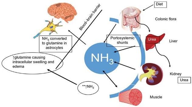
    8

    6

      • Hội chứng rối loạn ý thức & thay đổi hoạt động thần kinh cơ
      • Thường xảy ra ở trong suy tế bào gan cấp hoặc mạn hoặc có thông nối cửa chủ
      • Cơ chế bệnh sinh còn tranh cãi &

    có sự tham gia của nhiều chất trung gian

    Phân độ bệnh não gan

    • Độ I: thay đổi chu kì ngủ, hơi lú lẫn, dễ bị kích thích, run vẫy
    • Độ II: ngủ lịm, mất định hướng, thái độ bất thường, run vẫy
    • Độ III: lơ mơ, lú lẫn nặng, hung hăng, run vẫy
    • Độ IV: hôn mê

    Yếu tố thúc đẩy

    • Tăng urê huyết
    • Thuốc an thần, thuốc hướng tâm thần
    • Dẫn xuất á phiện
    • Xuất huyết tiêu hóa
    •  kali máu & kiềm máu (lợi tiểu, tiêu chảy)
    • Chế độ ăn nhiều đạm, bón
    • Nhiễm trùng
    • Rối loạn chức năng gan tiến triển
    • Thông nối cửa chủ (phẫu thuật, TIPS)

    Chẩn đoán

      • Biểu hiện đa dạng:

    Thay đổi tâm thần kinh nhẹ  hôn mê Run vẫy: bệnh não gan độ I-III,

    triệu chứng không đặc hiệu

      • EEG: sóng ba pha, chậm, biên độ cao
      • Định lượng NH3 máu

    không nhạy & không đặc hiệu

    Mục tiêu điều trị

    • Nhận biết & điều trị

    nguyên nhân & yếu tố thúc đẩy

    • Giảm sản xuất & hấp thu NH3 & các độc chất khác từ ruột:
      • giảm & thay đổi đạm trong chế độ ăn
      • thay đổi vi khuẩn đường ruột
      • thay đổi môi trường đường ruột
      • làm trống đường ruột
    • Thay đổi dẫn truyền thần kinh

    Điều trị yếu tố thúc đẩy

    • Tránh sử dụng thuốc an thần
    • Chống chỉ định Morphine, Paraldehyde
    • Không sử dụng acid amin dạng uống
    • Không dùng thuốc lợi tiểu
    • Bổ sung kali
    • Bổ sung kẽm
    • XHTH: cầm máu, loại bỏ máu

    Giảm sản xuất & hấp thu NH3

      • Chế độ ăn
      • Kháng sinh
      • Lactulose
      • Thụt tháo

    Chế độ ăn

    • Cơn cấp: đạm giảm còn 20 g/ngày
    • Lượng calo >25-35 kcal/kg: miệng, tĩnh mạch
    • Nếu hồi phục, tăng dần 10 g đạm /ngày
    • Nếu tái phát, trở lại mức điều trị trước
    • Đạm thực vật
      • nếu không dung nạp đạm động vật
      • ít sinh NH3, methionine, acid amin thơm
      • nhuận trường hơn & tăng lượng chất xơ
      • tăng sự hợp nhất & thải trừ nitơ qua phân
      • gây đầy hơi, tiêu chảy & nhiều phân

    Kháng sinh

    • Neomycin  sản xuất NH3 đường tiêu hóa Liều: 500-1000 mg mỗi 6 giờ

    thụt giữ 100-200 mL dung dịch 1% Thời gian: 5-7 ngày

    Phối hợp Lactulose có tác động hiệp lực 1-3% hấp thu, nguy cơ suy thận & độc tai

    Kháng sinh

      • Metronidazol 250 mg uống mỗi 6-8 giờ
        • hiệu quả như neomycin
        • độc tính trên hệ thần kinh trung ương
      • Rifaximin
        • không được hấp thu
        • hiệu quả đối với bệnh não gan độ 1-3
        • liều 400 mg uống 3 lần mỗi ngày
      • Vancomycin 250mg X 4 /ngày
        • sử dụng vancomycin khi kháng lactulose

    Lactulose

      • -1,4-galactosido-fructose – dissacharide
      • vi khuẩn ở ruột phân hủy thành a-xít lactic
      • giảm pH của phân, phân có tính a-xít
      • tăng khả năng thẩm thấu của đại tràng
      • tạo thuận lợi cho sự phát triển của vi khuẩn lên men lactose
      • ức chế vi khuẩn tạo NH3
      • giảm quá trình ion hóa & hấp thu NH3
      • rút ngắn diễn tiến bệnh của BNG sau XHTH

    Lactulose

    • Liều đầu: 15-45 ml uống 2-4 lần/ngày
    • Liều duy trì điều chỉnh để

    tiêu phân mềm 3-5 lần /ngày

    • Không sử dụng khi liệt ruột, tắc ruột
    • Tác dụng phụ
      • đầy hơi, tiêu chảy, đau bụng
      • tiêu chảy nặng:  Na,  K & kiềm máu
      • thể tích máu , suy thận

    Thụt tháo

    • Bệnh não gan do bón:

    giảm khi đi tiêu trở về bình thường

    • Dịch thụt tháo: trung tính hoặc có tính acid để làm  sự hấp thu NH3

    • Thụt tháo bằng lactulose tốt hơn nước 300 mL lactulose + 700 mL nước nhỏ giọt
    • Thụt tháo bằng MgSO4: tăng Mg máu
    • Thụt tháo với Phosphate an toàn hơn Mg

    Mục tiêu điều trị

    Nhận biết & điều trị

    nguyên nhân & yếu tố thúc đẩy

    Giảm sản xuất, hấp thu NH3 & các độc chất khác từ ruột

    • giảm & thay đổi đạm trong chế độ ăn
    • thay đổi vi khuẩn đường ruột
    • thay đổi môi trường đường ruột
    • làm trống đường ruột Thay đổi dẫn truyền thần kinh

    Thay đổi dẫn truyền thần kinh

    • Benzoate natri & L-ornithine-L-aspartate

      • Benzoate natri làm tăng bài tiết NH3 niệu

      • L-ornithine-L-aspartate thúc đẩy gan loại NH3 kích thích hoạt động chu trình urea gan thúc đẩy tổng hợp glutamine

    Thuốc: uống, tiêm tĩnh mạch

    →  nồng độ NH3 & cải thiện bệnh não

    Thay đổi dẫn truyền thần kinh

    • Levodopa & Bromocriptine Bệnh não có thông nối cửa chủ
      • Levodopa

    tiền thân của Dopamine gây tình trạng thức tỉnh

      • Bromocriptine

    chất đối vận thụ thể Dopamine đặc hiệu cải thiện khả năng tâm thần & EEG

    Thay đổi dẫn truyền thần kinh

    • Flumazenil
      • đối kháng thụ thể Benzodiazepine
      • cải thiện rõ tình trạng thần kinh & EEG
      • thời gian hoạt động rất ngắn

    Thay đổi dẫn truyền thần kinh

    • Các a-xít amin chuỗi ngắn Xơ gan

    Các AA chuỗi ngắn , các AA thơm 

    Tỉ lệ AA chuỗi ngắn / thơm 

    Truyền dịch AA chuỗi ngắn nồng độ cao

    → kết quả khác nhau, do khác biệt về: Thành phần của các dung dịch AA Cách sử dụng

    Đối tượng nghiên cứu

    Những biện pháp khác

    • Các phương pháp hỗ trợ gan tạm thời
      • phức tạp
      • không thích hợp với BNG do xơ gan
    • Ghép gan: trị liệu cuối cùng

    Thuốc và độc chất Rượu bia Methotrexate Amiodarone Vitamin A

    Bệnh do chuyển hóa/di truyền Nhiễm sắt

    Bệnh Wilson

    Thiếu alpha1-antitrypsin

    Viêm gan thoái hóa mỡ không do rượu

    Viêm gan virus ĐIỀU TRỊ NGUĐYa ÊnanNg đưNờnHg mÂậtNbẩm sinh

    Virus viêm gan B (±) D

    Virus viêm gan C

    Bệnh tự miễn

    Viêm gan tự miễn

    Xơ gan ứ mật tiên phát

    Teo hẹp đường mật

    GÂY XƠBGệnhAxơNhóa nang

    Bất thường về mạch máu Hội chứng Budd-Chiari Suy tim phải

    Viêm đường mật xơ hóa tiên phát

    Căn nguyên khác

    Bệnh gan thoái hóa hạt

    Hội chứng tắc nghẽn xoang

    Giãn mạch máu xuất huyết di truyền Xơ hóa tĩnh mạch cửa idiopathic

    88

    ĐIỀU TRỊ XƠ GAN DO RƯỢU

    EASL Clinical Practice Guidelines: Management of alcohol-related liver disease.

    Journal of Hepatology 2018 vol. 69 j 154–181

    89

    • Bệnh nhân xơ gan do rượu cần được khuyến cáo và khuyến khích để đạt được sự kiêng rượu hoàn toàn để giảm nguy cơ biến chứng và tử vong do gan
    • Xác định và điều trị các yếu tố đồng thời, như béo phì và kháng insulin, suy dinh dưỡng, hút thuốc lá, quá tải sắt và viêm gan virus
    • Sàng lọc và điều trị các biến chứng xơ gan nên được áp dụng đối với xơ gan do rượu

    VIÊM GAN B MẠN TRÊN XƠ GAN CÒN BÙ

    Xơ gan còn bù và lượng virus <2.000 IU/mL, nên được điều trị kháng virus để giảm nguy cơ mất bù, bất kể nồng độ ALT (AASLD 2018)

    1. Tenofovir và entecavir được ưu tiên vì hiệu lực và ít nguy cơ bị kháng thuốc, mất bù và tác dụng phụ nghiêm trọng. Thuốc kháng virus có hàng rào di truyền thấp với đề kháng không được khuyến cáo vì sự xuất hiện kháng thuốc có thể dẫn đến mất bù. TAF là thuốc kháng virus được ưu tiên bổ sung
    2. Peg‐IFN không chống chỉ định ở bệnh nhân xơ gan còn bù, nhưng NA an toàn hơn
    3. Nếu không điều trị, theo dõi tăng HBV DNA và/hoặc mất bù lâm sàng mỗi 3-6 tháng. Điều trị bắt đầu nếu xảy ra một trong hai

    90

    VIÊM GAN B MẠN TRÊN XƠ GAN CÒN BÙ

    1. ALT trong XG còn bù thường bình thường hoặc <2 lần so với giới hạn bình thường trên. ALT > 2 lần, tìm nguyên nhân khác  ALT và nếu không tìm thấy, thì đó là chỉ định mạnh hơn cho điều trị kháng virus
    2. Chứng cứ hiện tại không cung cấp thời gian điều trị tối ưu. Nếu ngừng điều trị, theo dõi ít nhất 1 lần/3 tháng/ ít nhất 1 năm, cho phép phát hiện sớm sự phát triển của virus có thể dẫn đến mất bù
    3. XG còn bù và HBV-DNA cao (>2.000 U/mL) được điều trị theo các khuyến nghị cho viêm gan virus B mạn HBeAg (-) và HBeAg (+)
    4. Điều trị thuốc kháng virus không loại trừ được nguy cơ HCC, cần tiếp tục theo dõi HCC

    91

    VIÊM GAN B MẠN TRÊN XƠ GAN MẤT BÙ

    XGMB HBsAg (+) được điều trị thuốc kháng virus vô thời hạn bất kể nồng độ HBV DNA, HBeAg hoặc ALT để giảm nguy cơ biến chứng do gan xấu đi (AASLD 2018)

    1. Entecavir và tenofovir là thuốc được khuyến cáo. TAF chưa được nghiên cứu trong XGMB, do đó hạn chế sử dụng TAF. TAF hoặc entecavir nên được xem xét ở bệnh nhân XGMB có rối loạn chức năng thận và/hoặc bệnh về xương
    2. Chống chỉ định Peg‐IFN ở XGMB do tính an toàn
    3. Cân nhắc ghép gan ở những người đủ điều kiện
    4. Theo dõi chặt chẽ để phát hiện tác dụng phụ của điều trị kháng virus: suy thận, nhiễm toan lactic
    5. Điều trị thuốc kháng virus không loại trừ được nguy cơ HCC, cần tiếp tục theo dõi HCC

    92

    Điều trị Viêm gan C (không hoặc có đồng nhiễm HIV) ở bệnh nhân xơ gan còn bù (Child-Pugh A) (EASL 2018)

    HCV Điều trị trước SOF/VEL GLE/PIB SOF/VEL

    /VOX

    SOF/LED GZR/EBR OBV/PIV/ r+DSV
    G1a Không 12 tuần 12 tuần không 12 tuần 12 tuần

    (≤8.105 IU/ml)

    không
    G1a 12 tuần 12 tuần không không không
    G1b K/Có 12 tuần 12 tuần không 12 tuần 12 tuần 12 tuần
    G2 K/Có 12 tuần 12 tuần không không không không
    G3 K/Có không 12 tuần 12 tuần không không không
    G4 Không 12 tuần 12 tuần không 12 tuần 12 tuần (≤8.105 IU/ml) không
    G4 12 tuần 12 tuần không không không không
    G5 Không 12 tuần 12 tuần không 12 tuần không không
    G5 12 tuần 12 tuần không không không không
    G6 Không 12 tuần 12 tuần không 12 tuần không không
    G6 12 tuần 12 tuần không không không không

    DSV: dasabuvir; EBR: elbasvir; GLE: glecaprevir; GZR: grazoprevir; LDV: ledipasvir; OBV: ombitasvir; PIB: pibrentasvir; PTV: paritaprevir; r: ritonavir; SOF: sofosbuvir; VEL: velpatasvir; VOX: voxilaprevir

    93

    94

    KẾT LUẬN

    Võ Thị Mỹ Dung [email protected] Bộ môn Nội tổng quát

    XG là giai đoạn cuối của nhiều loại bệnh gan mạn

    BN XGMB thường nhập viện & có nguy cơ cao bị tử vong do biến chứng

    Điều trị lý tưởng ở giai đoạn mất bù là ngăn ngừa xơ gan tiến triển

     

  • These slides give a comprehensive overview of the EASL clinical practice guidelines on the management of decompensated cirrhosis

    Clinical Practice Guidelines

    Decompensated

    cirrhosis

    These slides give a comprehensive overview of the EASL clinical practice guidelines on the management of decompensated cirrhosis

    Please feel free to use, adapt, and share these slides for your own personal use; however, please acknowledge EASL as the source

    • Definitions of all abbreviations shown in these slides are provided within the slide notes

    When you see a home symbol like this one: , you can click on this to return to the outline or topics pages, depending on which section you are in
    These slides are intended for use as an educational resource and should not be used in isolation to make patient management decisions. All information included should be verified before treating patients or using any therapies described in these materials

      • Paolo Angeli
    • Panel
      • Càndid Villanueva, Claire Francoz, Rajeshwar P Mookerjee, Jonel Trebicka, Aleksander Krag,

    Wim Laleman,

    Pere Gines,

    Mauro Bernardi (EASL Governing Board Representative)

    • Reviewers
      • Alexander Gerbes, Thierry Gustot, Guadalupe Garcia-Tsao

    Methods

    Background

    Guidelines

    Methods

    Grading evidence and recommendations

    Grading evidence and recommendations

    Grading is adapted from the GRADE system1

    Grade of evidence
    I Randomized, controlled trials
    II-1 Controlled trials without randomization
    II-2 Cohort or case-control analytical studies
    II-3 Multiple time series, dramatic uncontrolled experiments
    III Opinions of respected authorities, descriptive epidemiology
    Grade of recommendation
    1 Strong recommendation: Factors influencing the strength of the recommendation included the quality of the evidence, presumed patient-important outcomes, and cost
    2 Weaker recommendation: Variability in preferences and values, or more

    uncertainty: more likely a weak recommendation is warranted

    Recommendation is made with less certainty: higher cost or resource consumption

    1. Guyatt GH, et al. BMJ. 2008:336:924–6;


    EASL CPG decompensated cirrhosis. J Hepatol 2018;doi: 10.1016/j.jhep.2018.03.024

    Background

    Definition and pathophysiology of decompensated cirrhosis

    ACLF

    Stage 6: late decompensation:

    Refractory ascites, persistent PSE or jaundice,

    infections, renal and other organ dysfunction

    End stage

    Death

    • Transition from compensated cirrhosis to DC occurs at a rate of ~5–7% per year
    • DC is a systemic disease, with multi-organ/system dysfunction
    Compensated
    Stage 0: no varices, mild PH

    LSM >15 and <20 or HVPG >5 and <10 mmHg

    Stage 1: no varices, CSPH

    LSM ≥20 or HVPG ≥10 mmHg

    Stage 2: varices (=CSPH)
    Decompensated
    Stage 3: Bleeding
    Stage 4:

    First non-bleeding decompensation

    Stage 5:

    Second decompensating event

    • Transition from compensated cirrhosis to DC occurs at a rate of ~5–7% per year
    • DC is a systemic disease, with multi-organ/system dysfunction

    Stage 0: no varices, mild PH

    LSM >15 and <20 or HVPG >5 and <10 mmHg

    Stage 1: no varices, CSPH

    LSM ≥20 or HVPG ≥10 mmHg

    Stage 2: varices (=CSPH)

    Stage 3: Bleeding

    Stage 4:

    First non-bleeding decompensation

    Stage 5:

    Second decompensating event

    ACLF

    Stage 6: late decompensation:

    Refractory ascites, persistent PSE or jaundice,

    infections, renal and other organ dysfunction

    End stage

    Median survival: 2 years

    Symptomatic

    Decompensated

    Median survival: 12 years

    Asymptomatic

    Compensated

    Death

    Liver injury

    Cirrhosis

    Damaged cells/DAMPs

    Portal hypertension
    Bacterial translocation/PAMPs

    Other potential mechanisms

    Release of pro-inflammatory molecules (ROS/RNS)

    Activation of innate pattern recognition receptors

    ++

    Splanchnic arteriolar vasodilation and

    cardiovascular dysfunction

    Adrenal dysfunction

    HE Kidney

    dysfunction

    HPS

    Bernardi M, et al. J. Hepatol 2015;63:1272–84;

    • Management of DC aims to improve outcomes of complications

    Refractory

    Ascites

    Uncomplicated

    Hepatic hydrothorax

    CKD

    AKI

    Renal impairment

    Hyponatremia

    Complications of DC

    Bacterial infections

    ACLF

    RAI

    GI bleeding

    PPHT

    HPS

    CCM

    Cardiopulmonary

    Increased understanding of DC pathophysiology permits the development of more comprehensive therapeutic and prophylactic approaches

    to prevent or delay disease progression

    Key recommendations

    Overall management of DC

    Click on a topic to skip

    to that section

    1. Management of specific complications of DC
    • Management should aim to prevent progression, not treat complications

    No treatment exists that can act on cirrhosis progression directly

    • Two alternative approaches can be taken:
      • Suppress aetiological factor(s) that cause liver inflammation and cirrhosis development
      • Target key factors in the pathogenesis of cirrhosis decompensation and progression

    Impact is variable

    • Probably depends on the status of liver disease at the time
    Recommendation Grade of evidence Grade of recommendation
    In patients with DC the aetiological factor should be removed, particularly alcohol consumption and hepatitis B or C virus infection, as this strategy is associated with decreased risk of decompensation and increased survival II-2 1

    Several strategies have been evaluated to prevent disease progression in patients with DC

      • Targeting microbiome abnormalities and bacterial translocation to improve the gut–liver axis (i.e. rifaximin)
      • Improving the disturbed circulatory function (i.e. long-term albumin)
      • Treating the inflammatory state (i.e. statins)
      • Targeting portal hypertension (i.e. β-blockers)

    Further clinical research is needed to confirm the safety and potential benefits of these therapeutic approaches to

    prevent cirrhosis progression in patients with DC

    Most common complication of decompensation in cirrhosis

      • Develops in 5–10% of patients with compensated cirrhosis per year

    Significant impact on patients

      • Impairs patient working and social life
      • Frequently leads to hospitalization
      • Requires chronic treatment
      • Direct cause of further complications
      • Poor prognosis (5-year survival, ~30%)

    Ascites can be uncomplicated or refractory

      • Ascites is uncomplicated when not infected, refractory or associated with impairment of renal function

    Cirrhosis is responsible for 80% of cases of ascites

    • Initial patient evaluation:
      • History
      • Physical examination
      • Abdominal ultrasound
      • Laboratory assessment
        • Liver and renal function, serum and urine electrolytes, analysis of ascitic fluid

    Ascites is graded based on amount of fluid in the abdominal cavity

    Grading of ascites*
    Grade 1 Mild ascites: only detectable by ultrasound examination
    Grade 2 Moderate ascites: manifest by moderate symmetrical distension of abdomen
    Grade 3 Large or gross ascites: provokes marked abdominal distension

    *Ascites recurring on ≥3 occasions within a 12-month period despite dietary sodium restriction and adequate diuretic dosage are considered recurrent

    Diagnostic paracentesis is indicated in:*

      • All patients with new-onset grade 2 or 3 ascites
      • Patients hospitalized for worsening ascites or any complication of cirrhosis
    Recommendation Grade of evidence Grade of recommendation
    Neutrophil count and culture of ascitic fluid culture† should be

    performed to exclude bacterial peritonitis

    • Neutrophil count >250 cells/µl denotes SBP
    II-2 1
    Ascitic total protein concentration should be performed to identify patients at higher risk of developing SBP‡ II-2 1
    The SAAG should be calculated when the cause of ascites is not immediately evident, and/or when conditions other than cirrhosis are suspected§ II-2 1
    Cytology should be performed to differentiate malignancy-related from non-malignant ascites II-2 1

    *Grade of evidence II-2, grade of recommendation 1; †Bedside inoculation blood culture bottles with 10 ml fluid each;

    ‡A total protein concentration <1.5 g/dl is generally considered a risk factor for SBP;

    §SAAG ≥1.1 g/dl indicates that portal hypertension is involved in ascites formation with an accuracy of about 97%

    Development of ascites in patients with cirrhosis is associated with a poor prognosis

      • 1-year mortality: 40%
      • 2-year mortality: 50%

    Patients with ascites should be considered for referral for LT

    Recommendation Grade of evidence Grade of recommendation
    Since the development of grade 2 or 3 ascites in patients with cirrhosis is associated with reduced survival, LT should be considered as a potential treatment option II-2 1
    • Patients may not receive adequate priority in transplant lists

    – Most commonly used prognostic scores can underestimate mortality risk

    Improved methods to assess prognosis in these patients are needed

    • Grade 1 or mild ascites
      • No data on evolution and not known if treatment modifies natural history
    • Grade 2 or moderate ascites
      • Hospitalization not required
      • Correct sodium imbalance:
        • Dietary restriction and increased renal excretion with diuretics
    Recommendation Grade of evidence Grade of recommendation
    Moderate restriction of sodium intake (80–120 mmol/day,

    corresponding to 4.6–6.9 g of salt) is recommended

    I 1
    Generally equivalent to a no added salt diet with avoidance of

    pre-prepared meals. Adequate nutritional education of patients on how to manage dietary sodium is also recommended

    II-2 1
    Very low sodium diets (<40 mmol/day) should be avoided II-2 1
    Prolonged bed rest cannot be recommended III 1

    Mainstay of medical treatment are anti-mineralocorticoid drugs*

    • Loop diuretics may be added in patients with long-standing ascites
    Recommendation Grade of evidence Grade of recommendation
    First episode of grade 2 ascites

    • Anti-mineralocorticoid drug alone (from 100 mg/day with 100 mg stepwise increased every 72 hours to a maximum of 400 mg/day if no response to lower doses)
    I 1
    In patients who do not respond to anti-mineralocorticoids† or who develop hyperkalaemia, furosemide should be added (from 40 mg/day with 40 mg stepwise increases to a maximum of 160 mg/day) I 1
    Long-standing or recurrent ascites

    • Combination of an anti-mineralocorticoid drug and furosemide (dose increased sequentially according to response)
    I 1
    Torasemide can be given in patients exhibiting a weak response to furosemide I 2

    *Spironolactone, canrenone or K-canrenoate; †Body weight reduction <2 kg/week

    Patients with cirrhosis and ascites are highly susceptible to rapid reductions in extracellular fluid volume

      • Can lead to renal failure and hepatic encephalopathy
    Recommendation Grade of evidence Grade of recommendation
    GI haemorrhage, renal impairment, hepatic encephalopathy, hyponatraemia, or alterations in serum potassium concentration, should be corrected before starting diuretic therapy

    • In these patients, cautious initiation of diuretic therapy and frequent clinical and biochemical assessments should be performed
    III 1
    Diuretic therapy is generally not recommended in patients with

    persistent overt hepatic encephalopathy

    III 1

    Loop diuretics can lead to potassium and magnesium depletion and hyponatraemia

    • Muscle cramps can impair quality of life in patients receiving diuretics
    Recommendation Grade of evidence Grade of recommendation
    Frequent clinical and biochemical monitoring during the first weeks of treatment (particularly on first presentation) I 1
    Recommended maximum weight loss: 0.5 kg/day in patients without oedema, 1 kg/day in patients with oedema II-2 1
    Once ascites have largely resolved, the dose of diuretics should be

    reduced to the lowest effective dose

    III 1
    Discontinue diuretics in case of severe hyponatraemia,* AKI, worsening hepatic encephalopathy, or incapacitating muscle cramps III 1
    Discontinue furosemide for severe hypokalaemia (<3 mmol/L ) Discontinue anti-mineralocorticoids for hyperkalaemia (>6 mmol/L) III 1
    Albumin infusion or baclofen administration† are recommended in

    patients with muscle cramps

    I 1

    *Serum sodium <125 mmol/L; †10 mg/day, with a weekly increase of 10 mg/day up to 30 mg/day

    • Grade 3 or large ascites
      • LVP, under strict sterile conditions, is the treatment of choice
        • Ascites should be completely removed in a single session*
      • Contraindications to LVP include:
        • Uncooperative patient, abdominal skin infection at puncture sites, pregnancy, severe coagulopathy, severe bowel distention
    Recommendation Grade of evidence Grade of recommendation
    LVP should be followed with plasma volume expansion I 1
    Plasma volume expansion should be performed by albumin infusion (8 g/L ascites)

    • For >5 L of ascites: more effective than other plasma expanders
    • For <5 L of ascites (low risk of PPCD): treat with albumin due to concerns about use of alternative plasma expanders
    I III 1

    1

    After LVP, patients should receive the minimum dose of diuretics

    necessary to prevent re-accumulation of ascites

    I 1
    When needed, LVP should be performed in patients with AKI or SBP III 1

    *Grade of evidence I, grade of recommendation 1

    • Patients with DC and ascites are at increased risk of renal impairment from several types of drug
    Recommendation Grade of evidence Grade of recommendation
    NSAIDs should not be used (high risk of developing further sodium retention, hyponatraemia, and AKI) II-2 1
    Angiotensin-converting enyzme inhibitors, angiotensin II antagonists, or 1-adrenergic receptor blockers should not generally be used (increased risk of renal impairment) II-2 1
    Aminoglycosides are discouraged (increased risk of AKI)

    • Reserved for patients with severe bacterial infections that cannot be treated with other antibiotics
    II-2 1
    Contrast media

    • In patients with preserved renal function: does not appear to be associated with increased risk of renal impairment
    • In patients with renal failure: insufficient data, cautious use and preventative measures recommended
    II III 2

    1

    • International Ascites Club:

    “Ascites that cannot be mobilized or the early recurrence of which

    (after LVP) cannot be satisfactorily prevented by medical therapy”

    Diuretic intractable

    Diuretic resistant

    Refractory ascites

    Ascites that cannot be mobilized or the early recurrence of which cannot be prevented because of a lack of response to sodium

    restriction and diuretic treatment

    Ascites that cannot be mobilized or the early recurrence of which

    cannot be prevented because of the development of diuretic-induced complications that preclude the use of an effective diuretic dosage

    Diagnostic criteria
    Treatment duration Patients must be on intensive diuretic therapy* for at least 1 week and on a salt-restricted diet of less than 90 mmol/day
    Lack of response Mean weight loss of <0.8 kg over 4 days and urinary sodium output less than the sodium intake
    Early ascites recurrence Reappearance of grade 2 or 3 ascites within 4 weeks of initial mobilization
    Diuretic- induced complications
    • HE: development of encephalopathy in the absence of any other precipitating factor
    • Renal impairment: increase of serum creatinine by >100% to a value >2 mg/dl (177 µmol/L) in patients with ascites responding to treatment
    • Hyponatraemia: a decrease of serum sodium by >10 mmol/L to a serum sodium of <125 mmol/L
    • Hypo- or hyperkalaemia: a change in serum potassium to

    <3 mmol/L or >6 mmol/L despite appropriate measures

    • Incapacitating muscle cramps

    *Spironolactone 400 mg/day and furosemide 160 mg/day

    Refractory ascites is associated with a poor prognosis

      • Median survival around 6 months
    Recommendation Grade of evidence Grade of recommendation
    The diagnosis of refractory ascites relies on the assessment of the

    response of ascites to diuretic therapy and salt restriction

    • Evaluation should be done in stable patients without associated complications, such as bleeding or infection, after ascertaining patient compliance to treatment
    III 1
    Patients with refractory ascites should be evaluated for LT III 1

    LVP is a safe and effective treatment

      • Should be associated with albumin administration to prevent PPCD

    Drug treatments are controversial or inadequately studied

    Recommendation Grade of evidence Grade of recommendation
    Repeated LVP plus albumin (8 g/L of ascites removed) are recommended as first-line treatment for refractory ascites I 1
    Diuretics should be discontinued in patients with refractory ascites

    who do not excrete >30 mmol/day of sodium under diuretic treatment

    III 1
    Although controversial data exist on the use of NSBBs in refractory ascites, caution should be exercised in severe cases*

    • High doses of NSBB should be avoided (i.e. propranolol

    >80 mg/day)

    • Carvedilol can not be recommended at present
    II-2 I 1

    2

    *See also section on gastrointestinal bleeding

    TIPS decompresses the portal system*

      • Short term: accentuates peripheral arterial vasodilation
      • Within 4–6 weeks: improves effective volaemia and renal function to increase renal sodium excretion
    Recommendation Grade of evidence Grade of recommendation
    Patients should be evaluated for TIPS insertion when:

    • There is refractory or recurrent ascites
    • Paracentesis is ineffective
    I III 1

    1

    TIPS insertion is recommended in patients:

    • With recurrent ascites as it improves survival
    • With refractory ascites as it improves the control of ascites
    I

    I

    1

    1

    The use of small-diameter PTFE-covered stents is recommended to reduce the risk of TIPS dysfunction and hepatic encephalopathy I 1
    After TIPS insertion, continue the following until ascites resolution:

    • Diuretics and salt restriction
    • Close clinical follow-up
    II-2 III 1

    1

    *By shunting an intrahepatic portal branch into a hepatic vein

    Definition

      • Accumulation of transudate in the pleural space
        • In the absence of cardiac, pulmonary or pleural disease
      • Ascites moves through small diaphragmatic defects
        • Negative intrathoracic pressure induced by inspiration
      • Can lead to respiratory failure
      • Can be complicated by spontaneous bacterial infections (empyema)
      • Associated with poor prognosis
        • Median survival: 812 months

    Diagnosis

      • Once pleural effusion has been ascertained, cardiopulmonary and

    primary pleural diseases should be excluded*

      • Diagnostic thoracentesis is required to rule out bacterial infection*

    *Grade of evidence III, grade of recommendation 1

    • First-line management relies on treatment of ascites with diuretics and/or LVP
      • Not rare for pleural effusion to persist (refractory hepatic hydrothorax)

    Therapeutic thoracentesis is required to relieve dyspnoea

    Recommendation Grade of evidence Grade of recommendation
    Patients with hydrothorax should be evaluated for LT III 1
    Diuretics and thoracentesis are recommended as the first-line management of hepatic hydrothorax III 1
    Therapeutic thoracentesis is indicated in patients with dyspnoea III 1
    • Chronic pleural drainage should not be performed because of the
    frequent occurrence of complications II-2 1

    Hepatic hydrothorax:

    beyond diuretics and thoracentesis

    • Other treatments are appropriate in selected patients
    Recommendation Grade of evidence Grade of recommendation
    In selected patients, TIPS insertion for recurrent symptomatic hepatic hydrothorax is recommended II-2 1
    Pleurodesis can be suggested to patients with refractory hepatic hydrothorax not amenable to LT or TIPS insertion

    • The frequent occurrence of side effects restricts its use to selected patients
    I 2
    Mesh repair of diaphragmatic defects is suggested for the management of hepatic hydrothorax in very selected patients

    • The best results are achieved in patients with non-advanced cirrhosis without renal dysfunction
    II-2 2

    Hyponatraemia

    Common in patients with advanced cirrhosis

      • Arbitrarily defined as serum sodium concentration <130 mmol/L

    Hypo- and hypervolaemic hyponatraemia can occur

    • Associated with:
      • Increased mortality and morbidity, particularly neurological complications
      • Reduced survival after LT
    Recommendation Grade of evidence Grade of recommendation
    Patients with cirrhosis who develop hyponatraemia should be evaluated for LT II-2 1
    Removal of the cause and administration of normal saline are

    recommended in the management of hypovolaemic hyponatraemia

    III 1
    Fluid restriction* to 1,000 ml/day is recommended in the management of hypervolaemic hyponatraemia since it may prevent a further reduction in serum sodium levels III 1

    *Beyond fluid restriction, hypertonic saline should be limited to rare patients with life-threatening complications. It can be considered in patients with severe hyponatraemia who are expected to undergo LT within days. Correction of serum sodium concentration after attenuation of symptoms should be slow (≤8 mmol/L per day) to avoid irreversible neurological sequelae (II-3;1). Albumin can be administered but data are very limited (II-3;2). Use of vaptans should be limited to clinical trials (III;1)

    Occurs when variceal wall ruptures due to excessive wall tension

      • Portal pressure is a key factor in both rupture and severity of bleeding

    70% of GI bleeding events result from VH in patients with portal

    hypertension

      • Second most common decompensating event
      • Most severe and immediate life threatening complication
    Recommendation Grade of evidence Grade of recommendation
    Patients with DC are at high risk and should have an OGD to screen for varices, unless previously diagnosed and treated II-2 1
    If OGD is performed, the presence, size and presence of red wale marks should be reported II-2 1
    In patients without varices in whom aetiological factor persists and/or

    remain decompensated, screening OGD should be repeated yearly

    • In other patients the screening interval could be prolonged, but the exact interval is unclear and more data are required
    III 2

    High risk of death when VH occurs in patients with DC

      • Strategies to adequately treat VH and prevent (re)bleeding and death should be actively pursued
    Recommendation Grade of evidence Grade of recommendation
    Primary prophylaxis must be initiated upon detection of high-risk varices* because of increased risk of VH I 1
    Patients with small varices with red wale marks or Child–Pugh C

    should be treated with NSBBs

    III 1
    Patients with medium–large varices should be treated with either

    NSBBs or EBL

    • Choice of treatment can be based on local resources and expertise, patient preference, contraindications and AEs
    • NSBBs could be preferred because in addition to lowering portal

    pressure, they also exert other potential beneficial effects

    I III

    II-2

    1

    2

    2

    *High-risk = small varices with red signs, medium or large varices irrespective of Child–Pugh classification or small varices in Child–Pugh C patients

    NSBBs and EBL are equally effective in preventing first bleeding in patients with high-risk varices

      • Choice between options depends on factors such as patient preference, contraindications or adverse events
    Recommendation Grade of evidence Grade of recommendation
    Ascites is not a contraindication for NSBBs. However, caution should be exercised in cases of severe or refractory ascites

    • High doses of NSBBs should be avoided
    • The use of carvedilol can not be recommended at present
    I

    II-2 I

    1

    1

    2

    NSBBs should be discontinued in patients with progressive hypotension or those who develop an acute intercurrent condition* After recovery, reinstatement of NSBBs can be attempted

    • When NSBB intolerance or contraindications persist, patient’s bleeding risk should be managed by expeditious EBL
    III

    III III

    1

    2

    1

    NSBBs and EBL in combination reduces the risk of re-bleeding compared with monotherapy

    Recommendation
    Combination therapy of NSBBs + EBL is recommended I 1
    Covered TIPS placement is recommended in patients who continue to be intolerant to NSBBs* III 1

    *Provided that there are no absolute contraindications

    • Medical emergency: high rate of complications and mortality in DC
      • Requires immediate treatment and close monitoring

    Acute GI bleed + portal hypertension Initial assessment* and resuscitation

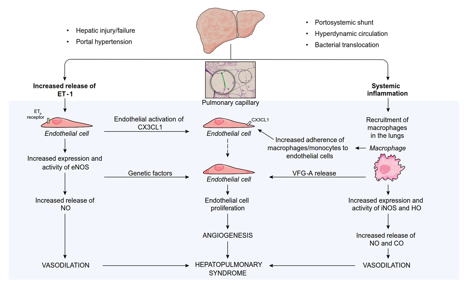
    Early diagnostic endoscopy (<12 hours)

    Confirm variceal bleeding

    Endoscopic band ligation

    Immediate start of vasoactive drug therapy† Antibiotic prophylaxis (I;1)‡

    Airway Breathing Circulation

    • Volume replacement with colloids and/or crystalloids should be initiated promptly (III;1)

    Starch should not be used (I;1)

    • Restrictive transfusion is recommended in most patients (Hb threshold, 7 g/dl; target range 7–9 g/dl) (I;1)

    ENDOSCOPY

    Balloon tamponade or oesophageal stenting

    (if massive bleeding)

    ENDOSCOPY

    +

    Maintain drug therapy for 3–5 days and antibiotics‡

    Control

    (~85% of cases)

    Further bleeding

    (~15% of cases)


    Consider early TIPS in high risk patients Rescue with TIPS

    *History, physical and blood exam, cultures; †Somatostatin/terlipressin; ‡Ceftriaxone (1 g/24 hours) is the first choice in patients with DC, those already on quinolone prophylaxis, and in hospital settings with high prevalence of quinolone-resistant bacterial infections. Oral quinolones (norfloxacin 400 mg BID) should be used in the remaining patients (I;1)

    Figure adapted from de Franchis R, et al. J Hepatol 2015;63:74352;

    Vasoactive drugs and ligation are the primary options for acute VH

      • There may be a role for TIPS in selected high-risk patients
    Recommendation Grade of evidence Grade of recommendation
    The combination of vasoactive drugs and ligation is recommended

    as the first therapeutic option in acute variceal bleeding

    I 1
    Early pre-emptive covered TIPS (placed within 24–72 hours) can be suggested in selected high-risk patients, such as those with Child–Pugh class C with score <14

    • However, the criteria for high-risk patients, particularly Child–Pugh B with active bleeding, remains debatable and needs further study
    I 2
    • Up to 10–15% of patients have persistent bleeding or early re-bleeding
      • Despite treatment with vasoactive drugs and EBL, and prophylactic antibiotics
    Recommendation Grade of evidence Grade of recommendation
    TIPS should be used as the rescue therapy of choice in cases of persistent bleeding or early re-bleeding I 1
    With the pre-requisite of expertise, balloon tamponade should be used in case of uncontrolled bleeding as a temporary ‘‘bridge” (max 24 hours) until definitive treatment can be instituted

    • Removable, covered and self-expanding oesophageal stents can be used as an alternative to balloon tamponade
    III

    I

    1

    2

    In the context of bleeding, where encephalopathy is commonly encountered, prophylactic lactulose may be used to prevent encephalopathy, but further studies are needed I 2
    β-blockers and vasodilators should be avoided during the acute

    bleeding episode

    III 1
    • Risk of bacterial infection in patients with cirrhosis is caused by multiple factors
      • Liver dysfunction
      • Portosystemic shunting
      • Gut dysbiosis
      • Increased BT
      • Cirrhosis-associated immune dysfunction
      • Genetic factors
    Spontaneous bacterial peritonitis
      • Definition: bacterial infection of ascitic fluid without any intra-abdominal surgically treatable source of infection
      • Prevalence: all patients with cirrhosis and ascites are at risk
        • 1.5–3.5% in outpatients; 10% in hospitalized patients
      • Prognosis: mortality exceeded 90% when first described
        • Reduced to ~20% with early diagnosis and treatment

    Diagnosis is based on diagnostic paracentesis

    • 50% of SBP episodes are present at hospital admission
      • Signs/symptoms of peritonitis: abdominal pain, tenderness, vomiting or

    diarrhoea, ileus

      • Signs of systemic inflammation: hyper- or hypothermia, chills, altered WBC count
      • Worsening liver function, HE, shock, renal impairment, GI bleeding

    However: SPB may be asymptomatic, particularly in outpatients

    Diagnosis is based on diagnostic paracentesis

    • 50% of SBP episodes are present at hospital admission
      • Signs/symptoms of peritonitis: abdominal pain, tenderness, vomiting or

    diarrhoea, ileus

      • Signs of systemic inflammation: hyper- or hypothermia, chills, altered WBC count
      • Worsening liver function, HE, shock, renal failure, GI bleeding
    Recommendation Grade of evidence Grade of recommendation
    Diagnostic paracentesis should be carried out in:

    • Patients with cirrhosis and ascites, at admission, to rule out SBP
    • Patients with GI bleeding, shock, fever or other signs of systemic inflammation, worsening liver and/or renal function, and HE
    II-2 1
    SBP diagnosed by a neutrophil count in ascitic fluid >250/mm3

    • Neutrophil count is determined by microscopy or flow cytometry
    • No clear evidence to support routine use of reagent strips
    II-2 1
    Ascitic fluid culture positivity is not a prerequisite for SBP diagnosis* II-2 1

    *Culture should be performed to guide antibiotic therapy (Grade of evidence II-2, grade of recommendation 1)

    • Empirical IV antibiotics should be started immediately following diagnosis*
    • Several factors should guide empirical antibiotic use†
      • Environment (community acquired vs. nosocomial)
      • Local bacterial resistance profiles
      • Severity of infection
    Recommendation Grade of evidence Grade of recommendation
    Third-generation cephalosporins are recommended as first-line antibiotic treatment for community-acquired SBP in countries with low rates of antibiotic resistance

    • In countries with high rates of antibiotic resistance

    piperacillin/tazobactam or carbapenem should be considered

    I

    II-2

    1

    1

    Antibiotic resistance is more likely in healthcare-associated and

    nosocomial SBP

    • Piperacillin/tazobactam: in areas with low prevalence of MDR bacteria
    • Carbapenem: in areas with high prevalence of ESBL-producing

    Enterobacteriaceae

    • Carbapenem + glycopeptides, daptomycin linezolid in areas with high prevalence of gram-positive MDR bacteria
    I 1

    *Grade of evidence II-2, grade of recommendation 1; †Grade of evidence I, grade of recommendation 1

    Antibiotic therapy should be carefully controlled and monitored

    Recommendation Grade of evidence Grade of recommendation
    Severe infections by XDR bacteria may require antibiotics known to be highly nephrotoxic in patients with cirrhosis (e.g. vancomycin or aminoglycosides)

    • In these cases, patients’ plasma levels should be monitored in

    accordance with local policy thresholds

    III 1
    De-escalation according to bacterial susceptibility based on positive cultures is recommended to minimize resistance selection pressure II-2 1
    Antibiotic efficacy should be checked with a second paracentesis at 48 hours from starting treatment

    • Suspect failure of first-line antibiotic if worsening clinical signs and symptoms and/or increase, or no marked reduction in leucocyte count (at least 25%) in 48 hours
    II-2 1
    The duration of treatment should be at least 5–7 days III 1
    Spontaneous bacterial empyema should be managed similarly to SBP II-2 2
    • In patients with SBP treated with a third generation intravenous cephalosporin antibiotic, albumin significantly decreased the incidence of type-1 hepatorenal syndrome and reduced mortality1
    Recommendation Grade of evidence Grade of recommendation
    The administration of albumin is recommended in patients with SBP

    • 1.5 g/kg at diagnosis and
    • 1 g/kg on Day 3
    I 1
    1. Sort P, et al. N Engl J Med 1999;341:403–9;

    Patients with cirrhosis and low ascitic fluid protein concentration (<10 g/L) and/or high serum bilirubin levels are at high risk of developing a first episode of SBP1

    Recommendation Grade of evidence Grade of recommendation
    Primary prophylaxsis with norfloxacin (400 mg/day) is recommended in patients with:

    • Child–Pugh score ≥9 and serum bilirubin level ≥3 mg/dl, and
    • Either impaired renal function or hyponatraemia, and
    • Ascitic fluid protein lower than 15 g/L
    I 1
    Norfloxacin prophylaxis should be stopped in patients with long-lasting improvement of their clinical condition and disappearance of ascites III 1
      • In patients who survive an episode of SBP, the cumulative recurrence rate at 1 year is approximately 70%1
    Recommendation Grade of evidence Grade of recommendation
    Prophylactic norfloxacin (400 mg/day, orally) is recommended in patients who recover from an episode of SBP I 1
    At present, rifaximin cannot be recommended as an alternative to

    norfloxacin for secondary prophylaxis of SBP

    • At present, no recommendation can be given to guide prophylaxis of SBP among patients already on rifaximin for the prevention of recurrent HE
    I 2
    Patients who recover from SBP have a poor long-term survival and

    should be considered for LT

    II-2 1
    PPIs may increase the risk for the development of SBP, their use

    should be restricted to those with a clear indication

    II-2 1

    Non-SBP infections are frequent in patients with cirrhosis

        • Associated with increased mortality
    Recommendation Grade of evidence Grade of recommendation
    Hospitalized patients with cirrhosis should be monitored closely

    for the presence of infections to enable early diagnosis and treatment

    II-1 1
    Empirical antibiotic therapy should be commenced promptly II-1 1
    Empirical antibiotic therapy should be based on: environment, local resistance profiles, severity and type of infection I 1
    In the context of high bacterial resistance to antibiotics, carbapenem alone or in combination with other antibiotics should be preferred* I 1
    Severe infections by XDR bacteria may require antibiotics known to be highly nephrotoxic in patients with cirrhosis (e.g. vancomycin or aminoglycosides)

    • In these cases, patients’ plasma level should be monitored in accordance with local policy thresholds
    III 1
    Routine use of albumin not recommended in infections other than SBP I 1

    *Carbapenem alone or in combination with other antibiotics proved to be superior to third-generation

    Good outcome

    qSOFA and Sepsis-3 criteria have been validated in patients with cirrhosis1

        • Can be used to assess severity of infection

    Is baseline SOFA score available?

    No

    Yes

    Apply sepsis-3 criteria and qSOFA

    Poor outcome

    Patient with need for transfer to ICU

    Sepsis-3 positive and qSOFA negative

    Good outcome

    Sepsis-3 and qSOFA positive

    Sepsis-3 and qSOFA negative

    Negative

    Positive

    Apply sepsis-3 criteria

    Grey zone Monitoring SOFA score is required

    1. Singer M, et al. JAMA 2016;315:801–10; Figure adapted from Piano S, et al. Gut 2017; doi: 10.1136/gutjnl-2017-314324.

    Nosocomial

    Healthcare- associated

    Community- acquired

    Other infections:

    recommended empirical antibiotic treatment

    Cellulitis

    Piperacillin- tazobactam or 3rd-gen

    cephalosporin + oxacillin

    AREA DEPENDENT:

    Like nosocomial infections if high prevalence of MDROs

    or if sepsis

    3rd-gen cephalosporin or meropemen + oxacillin or glycopeptides or daptomycin or linezolid

    Pneumonia UTI

    Nosocomial

    Healthcare- associated

    Community- acquired

    Nosocomial

    Healthcare- associated

    Community- acquired

    Piperacillin- tazobactam

    or ceftriaxone + macrolide or levofloxacin or moxifloxacin

    AREA DEPENDENT:

    Like nosocomial infections if high prevalence of MDROs

    or if sepsis

    Ceftazidime or meropemen + levofloxacin ± glycopeptides or linezolid

    UNCOMPLICATED:

    ciprofloxacin or cotrimoxazole

    IF SEPSIS: 3rd-gen cephalosporin

    or piperacillin-

    tazobactam

    AREA DEPENDENT:

    Like nosocomial infections if high prevalence of MDROs

    or if sepsis

    UNCOMPLICATED:

    fosfomycin or nitrofurantoin IF SEPSIS:

    meropemen + teicoplanin or vancomycin

    Other infections:

    recommended empirical antibiotic treatment

    Cellulitis

    Most clinically relevant

    3rd-gen cephalosporin or meropemen + oxacillin or glycopeptides or daptomycin or linezolid

    AREA DEPENDENT:

    Like nosocomial infections if high prevalence of MDROs

    or if sepsis

    Piperacillin- tazobactam or 3rd-gen

    cephalosporin + oxacillin

    Nosocomial

    Healthcare- associated

    Community- acquired

    UTI

    Pneumonia

    Ceftazidime or meropemen + levofloxacin ± glycopeptides or linezolid

    AREA DEPENDENT:

    Like nosocomial infections if high prevalence of MDROs

    or if sepsis

    Piperacillin- tazobactam

    or ceftriaxone + macrolide or levofloxacin or moxifloxacin

    Nosocomial

    Healthcare- associated

    Community- acquired

    Nosocomial

    Healthcare- associated

    Community- acquired

    UNCOMPLICATED:

    ciprofloxacin or cotrimoxazole

    IF SEPSIS: 3rd-gen cephalosporin

    or piperacillin-

    tazobactam

    AREA DEPENDENT:

    Like nosocomial infections if high prevalence of MDROs

    or if sepsis

    UNCOMPLICATED:

    fosfomycin or nitrofurantoin IF SEPSIS:

    meropemen + teicoplanin or vancomycin

    Definitions of renal impairment and staging of AKI have been updated

    Recommendation Grade of evidence Grade of recommendation
    In patients with liver diseases, even a mild increase in SCr should be considered since it may underlie a marked decrease of GFR II-2 1
    First step is to establish CKD, AKD, AKI or overlap II-2 1
    Diagnosis of CKD should be based on a GFR <60 ml/min/1.73 m2 estimated by SCr-based formulas, with or without signs of renal parenchymal damage* for at least 3 months II-2 1
    The diagnostic process should be completed by staging CKD, which

    relies on GFR levels, and by investigating its cause

    • Note that SCr-based formulae overestimate GFR in cirrhosis
    II-2 1
    Diagnosis of AKI should be based on adapted KDIGO criteria

    • Either an increase in SCr of >0.3 mg/dl from baseline within

    48 hours, or an increase of ≥50% from baseline within 3 months

    II-2 1
    Staging of AKI should be based on an adapted KDIGO system† II-2 1
      • Definitions of renal impairment and staging of AKI have been updated
    Recommendation Grade of evidence Grade of recommendation
    In patients with liver diseases, even a mild increase in SCr should be considered since it may underlie a marked decrease of GFR II-2 1
    First step is to establish CKD, AKD, AKI or overlap II-2 1
    Diagnosis of CKD should be based on a GFR <60 ml/min/1.73 m2 estimated by SCr-based formulas, with or without signs of renal parenchymal damage* for at least 3 months II-2 1
    The diagnostic process should be completed by staging CKD, which

    relies on GFR levels, and by investigating its cause

    • Note that SCr-based formulae overestimate GFR in cirrhosis
    II-2 1
    Diagnosis of AKI should be based on adapted KDIGO criteria

    • Either an increase in SCr of >0.3 mg/dl from baseline within

    48 hours, or an increase of ≥50% from baseline within 3 months

    II-2 1
    Staging of AKI should be based on an adapted KDIGO system† II-2 1

    KDIGO group definitions

    Definition Functional criteria Structural criteria
    AKI Increase in sCr ≥50% within 7 days,

    OR

    increase in sCr ≥0.3 mg/dl within 2 days

    No criteria
    AKD GFR <60 ml/min per 1.73m2 for <3 months,

    OR

    decrease in GFR ≥35% for <3 months,

    OR

    increase in sCr ≥50 % for <3 months

    Kidney damage

    for <3 months

    CKD GFR <60 ml/min per 1.73 m2 for ≥3 months Kidney damage for ≥3 months
      • KDIGO group definitions

    Increase in sCr ≥50% within 7 days,

    OR

    increase in sCr ≥0.3 mg/dl within 2 days

    Definition Functional criteria Structural criteria
    AKI Increase in sCr ≥50% within 3 months* No criteria
    AKD GFR <60 ml/min per 1.73m2 for <3 months,

    OR

    decrease in GFR ≥35% for <3 months,

    OR

    increase in sCr ≥50 % for <3 months

    Kidney damage

    for <3 months

    CKD GFR <60 ml/min per 1.73 m2 for ≥3 months Kidney damage for ≥3 months

    *International Club of Ascites-recommended adaptation of KDIGO group criteria

    Subject Definition
    Baseline sCr
    • sCr obtained within 3 months prior to admission
      • If >1 value within the previous 3 months, the value closest to the admission
    • If no previous sCr, the sCr on admission should be used
    Definition of AKI
    • Increase in sCr ≥0.3 mg/dl (≥26.5 µmol/L) within 48 hours or
    • Increase sCr ≥50% within the prior 7 days
    Staging of AKI
    • Stage 1: increase in sCr ≥0.3 mg/dl (≥26.5 µmol/L) or an increase in sCr ≥1.5-fold to 2-fold from baseline
    • Stage 2: increase in sCr >2-fold to 3-fold from baseline
    • Stage 3: increase of sCr >3-fold from baseline or sCr ≥4.0 mg/dl (353.6 µmol/L) with

    acute increase ≥0.3 mg/dl (≥26.5 µmol/L) or initiation of renal replacement therapy

    Progression of AKI Progression

    Progression of AKI to a higher stage and/or need for RRT

    Regression

    Regression of AKI to a lower stage

    Response to

    treatment

    No response No regression of AKI Partial response

    Regression of AKI stage with a reduction of sCr to ≥0.3 mg/dl (≥26.5 µmol/L) above baseline

    Full response

    Return of sCr to a value within

    0.3 mg/dl (≥26.5 µmol/L) of

    baseline

    Subject Definition
    Baseline sCr
    • sCr obtained within 3 months prior to admission
      • If >1 value within the previous 3 months, the value closest to the admission
    • If no previous sCr, the sCr on admission should be used
    Definition of AKI
    • Increase in sCr ≥0.3 mg/dl (≥26.5 µmol/L) within 48 hours or
    • Increase sCr ≥50% within the prior 7 days
    Staging of AKI Stage 1A (sCr <1.5mg/dl)*

    Stage 1B (sCr ≥1.5mg/dl)*

    • Stage 2: increase in sCr >2-fold to 3-fold from baseline
    • Stage 3: increase of sCr >3-fold from baseline or sCr ≥4.0 mg/dl (353.6 µmol/L) with

    acute increase ≥0.3 mg/dl (≥26.5 µmol/L) or initiation of renal replacement therapy

    Progression of AKI Progression

    Progression of AKI to a higher stage and/or need for RRT

    Regression

    Regression of AKI to a lower stage

    Response to

    treatment

    No response No regression of AKI Partial response

    Regression of AKI stage with a reduction of sCr to ≥0.3 mg/dl (≥26.5 µmol/L) above baseline

    Full response

    Return of sCr to a value within

    0.3 mg/dl (≥26.5 µmol/L) of

    baseline

    Stage 1: increase in sCr ≥0.3 mg/dl (≥26.5 µmol/L) or an increase in sCr ≥1.5-fold to 2- fold from baseline

    *ICA recommended adaptations of KDIGO group criteria

    Investigation and management should begin immediately

    Initial AKI* stage 1A

    Initial AKI* stage >1A

    NO

    YES

    Response

    Does AKI meet criteria of HRS?

    NO

    YES

    Specific treatment for

    other AKI phenotypes

    Vasoconstrictors and albumin

    *Initial AKI stage is defined as AKI stage at the time of first fulfilment of the AKI criteria;

    Persistance

    Further treatment of AKI decided on a case-by-case basics

    Close follow-up

    Progression

    Resolution

    Withdrawal of diuretics (if not yet applied) and volume expansion with albumin (1 g/kg) for 2 days

    Close monitoring

    Remove risk factors (withdrawal of nephrotoxic drugs, vasodilators and NSAIDs, taper/withdraw diuretics and β-blockers, expand plasma volume, treat infections† when diagnosed)

    †Treatment of spontaneous bacterial peritonitis should include albumin infusion according to current guidelines

      • All types of AKI can occur in patients with cirrhosis
        • Pre-renal, HRS, intrinsic, particularly ATN, and post-renal
      • Key point is to differentiate HRS-AKI from ATN
      • Classification of HRS was recently revised by the ICA1
        • Type 1 HRS now corresponds to HRS-AKI
        • Type 2 HRS includes renal impairment that fulfills the criteria of HRS but not of AKI (non-AKI-HRS or NAKI)
    Recommendation Grade of evidence Grade of recommendation
    It is important to differentiate among types of AKI in patients with

    cirrhosis

    II-2 1
    The diagnosis of HRS-AKI is based on revised ICA criteria (see next slide)

    • As kidney biopsy is rarely performed in the setting of AKI, biomarkers should be implemented
    • In clinical practice among the different biomarkers to date, urinary NGAL can be used to distinguish between ATN and HRS
    II-2 2
    1. Angeli et al. J. Hepatol 2015;62:968–74;
      • Cirrhosis and ascites
      • Diagnosis of AKI according to ICA-AKI criteria
      • No response after 2 consecutive days of diuretic withdrawal and plasma volume expansion with albumin 1 g per kg of body weight
      • Absence of shock
      • No current or recent use of nephrotoxic drugs (NSAIDs, aminoglycosides, iodinated contrast media, etc.)
      • No macroscopic signs of structural kidney injury,* defined as:
        • Absence of proteinuria (>500 mg/day)
        • Absence of microhaematuria (>50 RBCs per high power field)
        • Normal findings on renal ultrasonography

    *Patients who fulfil these criteria may still have structural damage such as tubular damage Angeli et al. J. Hepatol 2015;62:96874;

    First-line therapy is terlipressin plus albumin*

    Recommendation Grade of evidence Grade of recommendation
    All patients meeting the current definition of HRS-AKI stage >1A

    should be expeditiously treated with vasoconstrictors and albumin

    III 1
    Terlipressin can be administered by IV boluses (1 mg every 4–6 hours) or by continuous IV infusion (2 mg/day)†

    • In case of non-response (decrease in SCr <25% from the peak value) after 2 days, the dose of terlipressin should be increased in a stepwise manner to a maximum of 12 mg/day
    I 1
    Albumin solution (20%) should be used at 20–40 g/day

    • Serial measures assessing central blood volume can help to titrate the dose of albumin to prevent circulatory overload
    II-2 1
    Noradrenaline can be an alternative to terlipressin‡

    • Requires a central venous line often in an ICU Midodrine + octreotide can be an option when terlipressin or noradrenaline are unavailable (but efficacy is much lower)
    I I I 2

    1

    1

    *Grade of evidence I, grade of recommendation 1;

    †Continuous IV infusion allows for dose reduction to reduced adverse effects; ‡ Limited data are available

    Management of HRS-AKI: screening and monitoring

    Patients receiving treatment for HRS-AKI should be monitored for AEs and treatment response

        • AEs related to terlipressin or noradrenaline include ischaemic and cardiovascular events
    Recommendation Grade of evidence Grade of recommendation
    Careful clinical screening including ECG before starting the treatment is recommended

    • The decision to treat on a regular ward or transfer to higher dependency care should be case based

    Close monitoring of patients for the duration of treatment is important

    • Treatment should be modified or discontinued according to the

    type and severity of side effects

    I 1
    Response to treatment:

    • CR: final SCr within 0.3 mg/dl (26.5 µmol/L) from baseline
    • PR: regression of AKI stage to a final SCr ≥0.3 mg/dl (26.5 µmol/L) from baseline
    III 1
    In case of recurrence a repeat course of therapy should be given I 1

    *Continuous IV infusion allows for dose reduction to reduced adverse effects

    In most patients with HRS-AKI TIPS is contraindicated because of severe degree of liver failure

    Recommendation Grade of evidence Grade of recommendation
    There is insufficient data to advocate TIPS in HRS-AKI

    • It could be suggested in selected patients with HRS-NAKI
    II-2 2
    LT is the best therapeutic option for patients with HRS regardless of the response to drug therapy I 1
    The decision to initiate RRT should be based on the individual severity of illness I 2
    The indication for liver-kidney transplantation remains controversial

    • Should be considered in patients with significant CKD or sustained AKI including HRS-AKI with no response to drug therapy
    II-2 1
      • Based on the use of albumin in patients who develop SBP and the prevention of SBP using norfloxacin
    Recommendation Grade of evidence Grade of recommendation
    Albumin (1.5 g/kg at diagnosis and 1 g/kg on Day 3) should be given in patients with SBP to prevent AKI I 1
    Norfloxacin (400 mg/day) should be given as prophylaxis of SBP to prevent HRS-AKI I 1

    Frequent occurrence in cirrhotic patients

        • 30% of admitted patients and 25% of outpatients

    Major cause of death in patients with cirrhosis (50% mortality rate)

      • Develops on a background of acute decompensation

    Characterized by hepatic and extrahepatic organ failure, highly activated systemic inflammation and a high 28-day mortality

      • Precipitating events vary between populations and may include:
        • Bacterial infections (3057% of cases)
        • Active alcohol intake or alcohol binge
        • Reactivation of HBV
        • Superimposed HAV and HEV infection

    EASL-CLIF prognostic and diagnostic scores for ACLF

    10 x [0.033 x Clif OFs + 0.04 x Age + 0.63 x Ln(WBC) – 2]

    CLIF-C ACLF score for mortality prediction1*

    Chronic liver failure – organ failure score system1
    Organ/system† 1 point 2 points 3 points
    Liver (bilirubin, mg/dl) <6 ≥6–<12 ≥12.0
    Kidney (creatinine, mg/dl) <2.0 ≥2.0–<3.5 ≥3.5 or renal replacement
    Brain/HE (West Haven Criteria) Grade 0 Grades 1–2 Grades 3–4
    Coagulation (INR, PLT count) <2.0 ≥2.0–<2.5 ≥2.5
    Circulation (MAP, mmHg and

    vasopressors)

    ≥70 <70 Use of

    vasopressors

    Lungs PaO2/FiO2, or >300 ≤300–>200 ≤200§
    SpO2/FiO2 >357 >214–≤357 ≤214§

    *Age in years, creatinine in mg/dL, WBC in 106 cells/L, sodium in mmol/L;

    †Bold text indicates the diagnostic criteria for organ failures; ‡Patients submitted to mechanical ventilation due to HE and not to a respiratory failure were considered as presenting a cerebral failure (cerebral score = 3); §Other patients enrolled in the study with mechanical ventilation were considered as presenting a respiratory failure (respiratory score = 3)

    1. Jalan R, et al. J Hepatol 2014;61:1038–47;

    Even mild renal or brain dysfunction in the presence of another organ failure, is associated with a significant short-term mortality and therefore defines the presence of ACLF

    Grades of ACLF Clinical characteristics
    NO ACLF No organ failure, or single non-kidney organ failure, creatinine

    <1.5 mg/dl, no HE

    ACLF 1a Single renal failure
    ACLF 1b Single non-kidney organ failure, creatinine 1.5–1.9 mg/dl and/or HE grade 1–2
    ACLF II Two organ failures
    ACLF III Three or more organ failures

    Moreau R, et al. Gastroenterology 2013;144:1426–37;

    Diagnosis of ACLF is based on organ failure in the presence of AD in patients with cirrhosis

    Recommendation Grade of evidence Grade of recommendation
    ACLF diagnosis: cirrhosis and AD* plus organ failure(s) involving high short-term mortality II-2 1
    Diagnosis and grading should be based using the CLIF-C Organ Failure score II-2 1
    Potential precipitating factor(s) should be investigated†

    • Hepatic: heavy alcohol intake, viral hepatitis, DILI, autoimmune hepatitis
    • Extrahepatic: infections haemodynamic derangements following

    haemorrhage, surgery

    II-2 1

    *Defined as the acute development or worsening of ascites, overt encephalopathy, GI haemorrhage, non-obstructive jaundice and/or bacterial infections; †Note that in a significant proportion of patients, a precipitant factor may not be identified

    There is no specific therapy for ACLF

      • Treatment is based on organ support and management of

    complications

    Recommendation Grade of evidence Grade of recommendation
    Treatment of ACLF should be based on organ support, management

    of precipitants and associated complications

    III 1
    Patients should be treated in intermediate care or intensive care

    settings

    III 1
    ACLF is a dynamic condition and organ function should be

    monitored frequently and carefully throughout hospitalization

    • Particularly, liver, kidney, brain, lung, coagulation, and circulation
    • Monitoring and management should be individualized, mainly according to patients’ age and comorbidities
    III 1
      • The cause of liver injury can be treated in certain situations, e.g. HBV
      • Early action is crucial to patient survival
        • Treatment of precipitating factors
        • Referral for LT before evolution of ACLF makes LT impossible
    Recommendation Grade of evidence Grade of recommendation
    Early identification and treatment of precipitating factors of ACLF, particularly bacterial infections, is recommended.

    However, in some patients ACLF progresses despite treatment of

    precipitating factors

    III 1
    Nucleoside analogues (tenofovir, entecavir) should be instituted as

    early as possible in patients with HBV-related ACLF

    I 1
    Early referral of patients with ACLF to LT centres for immediate

    evaluation is recommended

    II-3 1
    Withdrawal of intensive care support after 1 week can be suggested

    in patients who are not LT candidates and have ≥4 organ failures

    II-2 2
    Administration of G-CSF cannot be recommended at present I 2

    Relative adrenal insufficiency

    • Inadequate cortisol response to stress in the setting of critical illness*

    – Pathophysiology in cirrhosis is not well defined

    • Diagnosis is influenced by the method used to measure cortisol
    • It is not known whether cortisol supplementation in clinically stable cirrhosis with RAI is of any value
    Recommendation Grade of evidence Grade of recommendation
    Diagnosis of RAI

    • <248 nmol/L (9 lg/dl) change in total serum cortisol after 250 lg corticotropin injection, or
    • Random total cortisol of <276 nmol/L (<10 lg/dl)
    II-2 1
    Salivary cortisol determination can be preferred

    • Serum free cortisol concentration can be influenced by reduced serum levels of CBG and albumin, frequently seen in patients with cirrhosis
    II-2 2
    Hydrocortisone treatment (at a dose of 50 mg/6 hours) of RAI

    cannot be recommended

    I 2

    *Also known as critical illness-related corticosteroid insufficiency (CIRCI)

    CCM occurs in patients with established cirrhosis characterized by:

        • Blunted contractile response to stress (pharmacological/surgery or inflammatory)
        • Altered diastolic left ventricular relaxation or/and increased left atrial volume
        • Electrophysiological abnormalities e.g. prolonged QTc
        • Cardiac output tending to decrease with decompensation
        • Systolic dysfunction: LVEF <55%

    CCM is largely subclinical but its presence influences prognosis in advanced disease

      • Numerous electrocardiographic criteria, along with transmitral Doppler assessment, are used for the evaluation and diagnosis of diastolic dysfunction
        • However, there is the need for more controlled studies and correlation with clinical endpoints
    Recommendation Grade of evidence Grade of recommendation
    ECG in patients with cirrhosis should be performed with dynamic stress testing* (systolic dysfunction may be masked by hyperdynamic circulation and reduced afterload)

    • Lack of increased CO after physiological/pharmacological stress† indicates systolic dysfunction
    II-1 1
    Myocardial strain imaging and assessment of GLS may be useful in

    the assessment of left ventricular systolic function in patients with DC

    II-2 2
    Cardiac MRI may identify structural changes III 2
    Diastolic dysfunction may occur as an early sign of CCM in the setting of normal systolic function, and should be diagnosed using ASE criteria:

    • Average E/e’>14
    • Tricuspid velocity >2.8 m/s
    • LAVI >34 ml/m2
    II-1 1

    *Either pharmacologically, or through exercise; †And in the absence of influence of β-blockade

      • Cardiac evaluation in patients with cirrhosis is important since CCM can influence prognosis
    Recommendation Grade of evidence Grade of recommendation
    In patients with AD, reduced CO (as a manifestation of CCM) is associated with the development of AKI (specifically hepatorenal dysfunction) after infections such as SBP II-1 1
    QTc interval prolongation is common in cirrhosis and may indicate a poor outcome

    • Agents that can prolong the QT interval should be used cautiously
    II-2 2
    Detailed functional cardiac characterization should be part of the assessment for

    • TIPS insertion
    • LT
    II-2 II-1 2

    1

    Standardized criteria and protocols for the assessment of systolic and

    diastolic function in cirrhosis are needed

    II-2 2
      • Four main pulmonary complications may occur in patients with chronic liver disease
        • Pneumonia
        • Hepatic hydrotorax
        • HPS
        • Portopulmonary hypertension
      • HPS is defined as a disorder in pulmonary oxygenation, caused by intrapulmonary vasodilatation and, less commonly, by pleural and pulmonary arteriovenous communications occurring in the clinical setting of portal hypertension
      • Clinical manifestations of HPS in patients with chronic liver disease primarily involve dyspnoea and platypnoea


    • Hepatic injury/failure
    • Portal hypertension
    • Portosystemic shunt
    • Hyperdynamic circulation
    • Bacterial translocation

    Increased ET-1 release

    ETB

    Endothelial activation

    receptor

    Pulmonary capillary

    CX3CL1

    Systemic inflammation

    Macrophage

    Endothelial cell

    of CX3CL1

    Genetic

    Increased eNOS

    expression and activity

    factors

    Endothelial cell

    Endothelial cell

    Increased adherence

    endothelial cells

    monocytes to

    of macrophages/

    VFG-A release

    recruitment

    in the lungs

    Endothelial cell

    proliferation

    Increased iNOS and HO

    expression and activity

    VASODILATION

    ANGIOGENESIS

    HEPATOPULMONARY SYNDROME

    Increased NO

    and CO release VASODILATION

      • Hypoxia with partial pressure of oxygen <80 mmHg or alveolar–arterial oxygen gradient ≥15 mmHg in ambient air (≥20 mmHg in patients older than 65 years)
      • Pulmonary vascular defect with positive findings on contrast-enhanced echocardiography or abnormal uptake in the brain (>6%) with radioactive lung-perfusion scanning
      • Commonly in presence of portal hypertension, and in particular:
        • Hepatic portal hypertension with underlying cirrhosis
        • Pre-hepatic or hepatic portal hypertension in patients without underlying cirrhosis
      • Less commonly in presence of:
        • Acute liver failure, chronic hepatitis
      • In patients with portal hypertension and the clinical suspicion of HPS partial pressure of oxygen (PaO2) in ABG should be assessed
    Recommendation Grade of evidence Grade of recommendation
    In patients with chronic liver disease, HPS should be suspected and investigated in presence of tachypnoea and polypnoea, digital clubbing and/or cyanosis II-2 1
    Screening in adults:

    • If pulse oximetry SpO2 <96% – ABG analysis should be performed
      • If ABG PaO2 <80mmHg and/or P[A-a]O2 ≥15 mmHg* (in ambient air) – further investigations should be performed
    II-2 1
    The use of contrast (microbubble) echocardiography to characterize HPS is recommended II-2 1

    *For adults ≥65 years a P[A-a]O2 ≥20 mmHg cut-off should be used

      • When PaO2 suggests HPS, further investigations are needed to determine the underlying mechanism
    Recommendation Grade of evidence Grade of recommendation
    MAA scan should be performed to quantify the degree of shunting in patients with severe hypoxaemia and coexistent intrinsic lung disease, or to assess the prognosis in patients with HPS and very severe hypoxaemia (PaO2 <50 mmHg) II-2 1
    Neither contrast echocardiography nor MAA scan can definitively differentiate discrete arteriovenous communications from diffuse precapillary and capillary dilatations or cardiac shunts

    • Pulmonary angiography should be performed only in patients with the severe hypoxaemia (PaO2 <60 mmHg), poorly responsive to administration of 100% oxygen, and in whom there is a strong suspicion of arteriovenous communications that are amenable to embolization
    II-2 1
    Trans-oesophageal contrast-enhanced echocardiography (although associated with risks) can definitively exclude intra-cardiac shunts II-2 2
      • There is no established medical therapy currently available for HPS, the only successful treatment for HPS is LT
    Recommendations for medical treatment Grade of evidence Grade of recommendation
    Long-term oxygen therapy is recommended in patients with HPS and severe hypoxaemia despite the lack of available data concerning effectiveness, tolerance, cost effectiveness, compliance and effects on survival rates of this therapy II-2 1
    No recommendation can be proposed regarding the use of drugs or the placement of TIPS for the treatment of HPS I 1
    Recommendations for liver transplantation
    Patients with HPS and PaO2 <60 mmHg should be evaluated for LT since it is the only treatment for HPS that has been proven to be effective to date II-2 1
    Severe hypoxaemia (PaO2 <45–50 mmHg) is associated with

    increased post-LT mortality

    • ABG analysis should be carried out every 6 months to facilitate prioritization to LT
    II-2 1
      • PPHT occurs in patients with portal hypertension in the absence of other causes of arterial or venous hypertension
      • Classification is based on mean pulmonary arterial pressure (mPAP), and assumes high pulmonary vascular resistance (PVR) and normal pulmonary occlusion pressures
        • Mild: mPAP ≥25 and <35 mmHg
        • Moderate: mPAP ≥35 and <45 mmHg
        • Severe: mPAP ≥45 mmHg
      • Incidence between 3–10% cirrhosis patients based on haemodynamic criteria; women are at 3x greater risk and it is more common in autoimmune liver disease
      • There is no clear association between the severity of liver disease or portal hypertension and the development of severe PPHT

    The evidence base for pharmacological therapies in PPHT is limited

    Recommendation Grade of evidence Grade of recommendation
    Screening for PPHT should be via TDE in patients deemed

    potential recipients for TIPS or LT

    • In those with a positive screening test, right heart catheterization should be performed
    II-1 1
    In patients with PPHT who are listed for LT, echocardiography should be repeated on the waitlist (the specific interval is unclear) III 1
    β-blockers should be stopped and varices managed by endoscopic therapy in cases of proven PPHT II-3 1
    Therapies approved for primary pulmonary arterial hypertension may improve exercise tolerance and haemodynamics in PPHT

    • However, endothelin antagonists should be used with caution

    because of concerns over hepatic impairment

    II-2 1
    TIPS should not be used in patients with PPHT II-3 1
      • Although severe PPHT has, historically, been a contraindication for LT, the advent of improved haemodynamic control (with agents such as IV prostacyclin) allows LT to be considered
    Recommendation Grade of evidence Grade of recommendation
    If mPAP <35 mmHg and right ventricular function is preserved, LT

    should be considered

    • mPAP of ≥45 mmHg should be considered an absolute contraindication to LT irrespective of therapy applied
    II-2 III 1

    1

    Therapy to lower mPAP and improve right ventricular function

    should be commenced in patients with mPAP ≥35 mmHg

    • Right ventricular function should be periodically evaluated
    II-2 1
    MELD exception can be considered in patients with proven PPHT in whom targeted therapy fails to decrease mPAP <35 mmHg but does facilitate normalization of PVR to <240 dyn.s/cm-5 and right ventricular function II-3 2
    MELD exception should be advocated in patients with proven PPHT of moderate severity (mPAP ≥35 mmHg) in whom targeted treatment lowers mPAP <35 mmHg and PVR <400 dyn.s/cm-5 II-2 1

    Additional recommendations

    Portal hypertension gastropathy

      • Often presents in patients with DC
        • Natural history significantly influenced by the severity of liver disease and portal hypertension
    Recommendation Grade of evidence Grade of recommendation
    NSBB and iron supplementation and/or blood transfusion, when indicated, are recommended as first-line therapy for chronic haemorrhage from PHG I 1
    In patients with transfusion-dependent PHG in whom NSBBs fail or are not tolerated, covered TIPS placement may be used in the absence of contraindications II-3 2
    Acute PHG bleeding may be treated with somatostatin analogues or terlipressin but substantiating data are limited I 2

    Gastric varices: classification, prevalence and risk

      • The Sarin classification is most commonly used for risk stratification and management of gastric varices
    Type Definition Relative frequency Overall bleeding risk without treatment
    Gastro-oesophageal varices (GOV)
    GOV type 1 OV extending below cardia into lesser curvature 70% 28%
    GOV type 2 OV extending below cardia into fundus 21% 55%
    Isolated gastric varices (IGV)
    IGV type 1 Isolated varices in the fundus 7% 78%
    IGV type 2 Isolated varices else in the stomach 2% 9%
      • Gastric varices are present in about 20% of patients with cirrhosis
    Recommendation Grade of evidence Grade of recommendation
    NSBBs are suggested for primary prevention of VH from GOV type 2 or IGV type 1 III 2
    Primary prevention for GOV type 1 follows the recommendations of oesophageal varices III 2
    Acute gastric VH should be treated medically, like oesophageal VH

    • Cyanoacrylate is the recommended endoscopic haemostatic treatment for cardiofundal varices (GOV type 2 or IGV type 1)
    I

    I

    1

    2

    TIPS with potential embolization efficiently controls bleeding and prevents re-bleeding in fundal VH (GOV type 2 or IGV type 1) and should be considered in appropriate candidates II-2 1
    Selective embolization (BRTO/BATO) may also be used to treat bleeding from fundal varices associated with large gastro/splenorenal collaterals, although more data is required III 2

    Both neutrophil count and culture results should be taken into account

    Recommendation Grade of evidence Grade of recommendation
    Patients with bacterascites (neutrophil count <250/mm3 but positive bacterial culture) exhibiting signs of systemic inflammation or infection should be treated with antibiotics

    • Otherwise, the patient should undergo a second paracentesis
      • If the culture results come back positive again, regardless of

    the neutrophil count, the patient should be treated

    II-2

    III

    1

    1

    Spontaneous bacterial pleural empyema diagnosed by:

    • Positive pleural fluid culture and neutrophil count >250/mm3 or
    • Negative pleural fluid culture and neutrophil count >500/mm3 in the absence of pneumonia
    II-2 1
    Secondary bacterial peritonitis should be suspected in case of multiple organisms on ascitic culture, very high ascitic neutrophil count and/or high ascitic protein concentration, or in those patients with an inadequate response to therapy

    • Patients with suspected secondary bacterial peritonitis should undergo prompt CT scanning and early considerations for surgery
    III 1
    SBP or SBE

    Nosocomial SBP or SBE

    Healthcare-associated SBP or SBE

    Community-acquired SBP or SBE

    3rd-gen cephalosporin or

    piperacillin-tazobactam

    AREA DEPENDENT:

    Like nosocomial infections if high prevalence of MDROs or if sepsis

    Carbapenem alone or + daptomycin, vancomycin (or linezolid*) if high prevalence of MDR Gram+ bacteria or sepsis

    *In areas with a high prevalence of vancomycin-resistant enterococci Adapted from Jalan R, et al. J Hepatol 2014;60:1310–24;

    Investigation and management should begin immediately

    Recommendation Grade of evidence Grade of recommendation
    Investigate AKI cause as soon as possible to prevent AKI progression

    • Management should begin immediately (even absent obvious cause)
    • Screening and treatment of infection are most important
    II-2 1
    Diuretics and/or β-blockers as well as other drugs that could be associated with the occurrence of AKI such as vasodilators, NSAIDs and nephrotoxic drugs should be immediately stopped II-2 1
    Volume replacement should be used in accordance with the cause and severity of fluid losses II-2 1
    In case of no obvious cause of AKI, AKI stage >1A or infection-induced AKI:

    • 20% albumin solution should be used at a dose of 1 g /kg of body weight (maximum of 100 g of albumin) for 2 consecutive days
    III 1
    In patients with AKI and tense ascites, therapeutic paracentesis should be associated with albumin infusion even when a low volume of ascitic fluid is removed III 1
      • HRS-NAKI has an impaired response to vasorestrictors
    Recommendation Grade of evidence Grade of recommendation
    Vasoconstrictors and albumin are not recommended the treatment of HRS outside the criteria of AKI (HRS-NAKI)*

    • Terlipressin plus albumin is effective in the treatment of

    HRS-NAKI, but recurrence after withdrawal of treatment is the norm, and controversial data exist on the impact of the treatment on long-term clinical outcome, particularly from the perspective of LT

    I 1

    *Formerly known as HRS type II

    Previous CLIF prognostic and diagnostic scores

    10 x [0.03 x Age + 0.66 x Ln(Creatinine) + 1.71 x Ln(INR) + 0.88 x

    Ln(WBC) 0.05 x Sodium + 8]

    CLIF-C Acute decompensation score1*

    Sequential Organ Failure Assessment (SOFA) score
    Organ/system* 0 1 2 3 4
    Liver (bilirubin mg/dl) <1.2 ≥1.2–<2.0 ≥2.0–<6.0 ≥6.0–<12.0 ≥12.0
    Kidney (creatinine mg/dl) <1.2 ≥1.2–<2.0 ≥2.0–<3.5 ≥3.5–<5.0 ≥5.0
    Cerebral (HE grade) NO HE Grade I Grade II Grade III Grade IV
    Coagulation

    (INR and PLT count)

    <1.1 ≥1.1–<1.25 ≥1.25–<1.5 ≥1.5–<2.5 ≥2.5 or PLT

    ≤20,000/mm3

    Circulation

    (MAP mmHg

    and vasopressors†)

    ≥70 <70 Dopamine ≤5 or dobutamine or terlipressin Dopamine >5

    or A ≤0.1 or NA ≤0.1

    Dopamine >15

    or A >0.1

    or NA >0.1

    Lungs
    PaO2/FiO2, or >400 >300–≤400 >200–≤300 >100–≤200 ≤100
    SpO2/FiO2 >512 >357–≤512 >214–≤357 >89- ≤214 ≤89

    *Bold text indicates the diagnostic criteria for organ failures; †μg/kg/min


  • TRIỆU CHỨNG, NGUYÊN NHÂN, CHẨN ĐOÁN VÀ ĐIỀU TRỊ LOÉT DẠ DÀY – LOÉT TÁ TRÀNG

    Dàn bài

      1. Đại cương
      2. Bệnh sinh
      3. Nguyên nhân
      4. Biểu hiện lâm sàng
      5. Cận lâm sàng
      6. Biến chứng
      7. Phòng ngừa

    ĐẠI CƯƠNG

    Loét dạ dày hoặc/và loét tá tràng là những vết loét ở niêm mạc dạ dày hoặc tá tràng, sâu xuống lớp cơ niêm. Cả hai bệnh loét dạ dày và loét tá tràng đều có liên quan đến tác động phá hủy niêm mạc của pepsin và a-xít hydrochloric ở đường tiêu hóa trên. Các ổ loét thường có đường kính từ 3 mm đến vài xen-ti-mét.

    Trong trường hợp bệnh Loét dạ dày-tá tràng không có biến chứng, có rất ít dấu hiệu lâm sàng và không đặc hiệu. Đau thượng vị là triệu chứng thường gặp nhất trong loét dạ dày và trong loét tá tràng.

    Các thử nghiệm cận lâm sàng thường qui thường không có ích đối với phần lớn bệnh nhân bị Loét dạ dày-tá tràng không có biến chứng. Nội soi đường tiêu hóa trên là thử nghiệm chẩn đoán được chọn.

    Dịch tễ học

    Ở Mỹ, hàng năm, Loét dạ dày-tá tràng ảnh hưởng đến khoảng 4,5 triệu người. Khoảng 10% dân số Mỹ có bằng chứng loét tá tràng tại một thời điểm bất kỳ. Nhìn chung, tỉ lệ mới bị loét tá tràng giảm trong 3-4 thập niên qua. Mặc dù, tỉ lệ loét dạ dày không có biến chứng giảm, nhưng tỉ lệ loét dạ dày có biến chứng và nhập viện vẫn không thay đổi. Tỉ lệ nhập viện vì Loét dạ dày-tá tràng khoảng 30 bệnh nhân trong 100.000 ca bệnh. Tỉ lệ bệnh Loét dạ dày-tá tràng thay đổi từ chiếm ưu thế ở nam thành tỉ lệ tương tự giữa nam và nữ. Tỉ lệ mắc bệnh trong suốt cuộc đời khoảng 11-14% ở nam và 8-11% ở nữ. Các xu thế tuổi bị loét giảm ở nữ trẻ, nhất là đối với loét tá tràng và tăng ở nữ lớn tuổi. Xu thế này phản ánh những thay đổi phức tạp về yếu tố nguy cơ của Loét dạ dày-tá tràng, gồm đoàn hệ tuổi với nhiễm H.pylori và sử dụng NSAIDs trong dân số người lớn tuổi. Ở những người nhiễm H.pylori, tỉ lệ bị bệnh Loét dạ dày-tá tràng trong suốt cuộc đời khoảng 20%. Chỉ khoảng 10% người trẻ bị nhiễm H.pylori; tỉ lệ nhiễm trùng ngày càng tăng theo tuổi.

    Tần suất Loét dạ dày-tá tràng ở các nước khác thay đổi.

    Giải phẫu

    Xem lại trong sách giải phẫu học

    BỆNH SINH

    Trước đây người ta cho rằng “No gastric acid, no peptic ulcer”. Tuy nhiên, tiết a- xít dạ dày quá nhiều chỉ là một yếu tố trong bệnh sinh của bệnh Loét dạ dày-tá tràng. Khả năng bảo vệ niêm mạc chống lại tác động phá hủy của a-xít dạ dày bị giảm cũng là căn nguyên gây loét. Tính toàn vẹn của đường tiêu hóa trên phụ thuộc vào tình trạng cân bằng giữa các yếu tố “phá hủy” như a-xít của dạ dày, H.pylori, NSAIDs và pepsin và các yếu tố “bảo vệ” như prostaglandins, chất nhầy, bicarbonate và lưu lượng máu đến niêm mạc ảnh hưởng đến niêm mạc dạ dày ruột.

    Bình thường, tế bào biểu mô của dạ dày hoặc tá tràng tiết chất nhầy chống lại những kích thích ở lớp biểu mô và kích thích cholinergic. Bề mặt niêm mạc dạ dày và tá tràng được phủ một lớp gel, lớp này không thấm a-xít và pepsin. Những tế bào nhầy của dạ dày và tá tràng tiết bicarbonate, bicarbonate giúp đệm lớp a-xít nằm kề cận niêm mạc. Prostaglandins E (PGE) có vai trò quan trọng trong bảo vệ niêm mạc dạ dày, vì PGE làm tăng sản xuất bicarbonate và chất nhầy.

    Trong trường hợp a-xít và pepsin đi vào tế bào biểu mô, có những cơ chế hỗ trợ để giảm bớt tổn thương. Bơm ion trong màng của các tế bào biểu mô giúp điều hòa pH trong tế bào bằng cách loại bỏ các ion hydrogen. Do tiến trình phục hồi, những tế bào lành sẽ di chuyển đến vị trí tổn thương. Dòng máu ở niêm mạc loại bỏ a-xít khuếch tán vào niêm mạc bị tổn thương và cung cấp bicarbonate cho tế bào biểu mô bề mặt. Trong điều kiện bình thường, cân bằng sinh lý giữa tiết a- xít dạ dày và bảo vệ niêm mạc dạ dày tá tràng.

    Tổn thương niêm mạc dạ dày và tá tràng xảy ra khi tác động có hại của a-xít dạ dày lấn át tính chất bảo vệ niêm mạc. Ức chế tổng hợp prostaglandin nội sinh làm giảm tiết bicarbonate, nhầy của tế bào biểu mô, giảm lưu lượng máu đến niêm mạc, giảm tăng sinh tế bào biểu mô và sức đề kháng của niêm mạc với chấn thương. Sức đề kháng của niêm mạc giảm khiến niêm mạc dễ bị tổn thương bởi những yếu tố nội sinh như a-xít, pepsin và a-xít mật và những yếu tố ngoại sinh như NSAIDs, rượu và các chất độc hại khác.

    Như vậy, tổn thương niêm mạc và Loét dạ dày-tá tràng xảy ra khi tình trạng cân bằng giữa các yếu tố phá hủy và cơ chế bảo vệ bị phá vỡ. Những yếu tố phá hủy như NSAIDs, nhiễm H.pylori, rượu bia, muối mật, a-xít và pepsin có thể làm thay đổi khả năng bảo vệ niêm mạc cho phép các ion hydrogen khuếch tán ngược và làm tổn thương tế bào biểu mô. Những cơ chế bảo vệ gồm tình trạng liên kết chặt chẽ giữa các bào, chất nhầy, dòng máu của niêm mạc, quá trình phục hồi tế bào và tình trạng tái sinh biểu mô.

    NGUYÊN NHÂN

    A-xít dạ dày

    1/3 bệnh nhân bị loét tá tràng tăng cung lượng a-xít cơ bản (BAO) và cung lượng a-xít tối đa (MAO). Trong một nghiên cứu, BAO tăng đi kèm với tỉ số chênh 3,5 và MAO tăng đi kèm với tỉ số chênh 7 đối với sự phát triển loét tá tràng. Những người đặc biệt có nguy cơ cao là những người có BAO lớn hơn 15 mEq/giờ.

    H.pylori

    Mối liên hệ giữa xoắn khuẩn H.pylori gram âm với viêm dạ dày được đề cập lần đầu tiên vào năm 1983. Từ đó, các nghiên cứu phát hiện H.pylori là một phần quan trọng trong bộ ba bao gồm a-xít và pepsin, góp phần khởi đầu gây bệnh Loét dạ dày-tá tràng. Vi khuẩn gây viêm niêm mạc và ở một số người, vi khuẩn làm bệnh loét dạ dày trầm trọng hơn. H.pylori có những đặc điểm vi sinh độc đáo, như sản xuất urease làm kiềm hóa môi trường xung quanh vi khuẩn và tồn tại trong nhiều năm trong môi trường a-xít không thân thiện của dạ dày và sản xuất những yếu tố độc lực khác như catalase, cytotoxin tạo không bào và lipopolysaccharide.

    H.pylori định cư ở niêm mạc dạ dày, thường gây viêm niêm mạc. Mối liên hệ nhân quả giữa viêm dạ dày do H.pylori và loét tá tràng hiện nay đã được chứng minh rõ trong y văn trên người. Ở những bệnh nhân bị nhiễm H.pylori, nồng độ gastrin và pepsinogen cao, nồng độ somatostatin bị giảm và tá tràng tăng tiếp xúc với a-xít. Ngoài ra, ở bệnh nhân bị loét tá tràng, thường làm trống dạ dày nhanh; một lượng lớn a-xít tống thoát vào đoạn đầu tá tràng, do đó loét hành tá tràng chiếm 95% loét tá tràng. Hầu hết ở bệnh nhân loét tá tràng, bài tiết bicarbonate của tá tràng bị hư hỏng, điều này đã được chứng minh là do H.pylori vì tiệt trừ H.pylori làm thay đổi hoàn toàn khiếm khuyết này. Tăng tiết a-xít dạ dày và giảm tiết bicarbonate tá tràng càng làm giảm thấp pH ở tá tràng. Toan hóa tá tràng khiến chuyển sản dạ dày – thay thế các tế bào có lông của tá tràng bằng những tế bào có đặc điểm về hình dạng và bài tiết của biểu mô dạ dày – tạo môi trường thích hợp cho H.pylori định cư. Nhiễm H.pylori trong những vùng chuyển sản dạ dày gây viêm tá tràng và khiến niêm mạc dễ nhạy cảm với tổn thương do a-xít, tạo điều kiện thuận lợi gây loét tá tràng.

    Nhiễm trùng H.pylori ở dạ dày giải thích cho đa số các trường hợp Loét dạ dày- tá tràng. Vi khuẩn này cũng có vai trò trong sự hình thành lymphoma dạng MALT và ung thư biểu mô tuyến dạ dày. Nhiễm trùng H.pylori thực sự có liên quan với viêm dạ dày hoạt động, nhưng chỉ khoảng 10-15% người bị nhiễm trùng phát triển thành Loét dạ dày-tá tràng thực sự. Tác động của H.pylori trên đường tiêu hóa (Viêm dạ dày, Loét dạ dày-tá tràng, Lymphoma dạng MALT, Ung thư dạ dày) là hậu quả của tác động tương hỗ phức tạp giữa kí chủ và vi khuẩn.

    Yếu tố vi khuẩn

    H.pylori có thể tự tạo điều kiện thuận lợi để cư trú ở dạ dày, gây tổn thương niêm mạc dạ dày và thoát khỏi hệ thống bảo vệ của ký chủ. Các chủng H.pylori khác nhau sản xuất yếu tố độc tính khác nhau. Vùng đặc hiệu của bộ gen của vi khuẩn, còn được gọi là đảo gây bệnh, mã hóa các yếu tố độc tính Cag A và pic

    B. Mặc dù không được mã hóa trong đảo gây bệnh, nhưng Vac A cũng góp phần sinh bệnh. Những yếu tố độc lực này, cùng với những yếu tố cấu thành của vi khuẩn, có thể gây tổn thương niêm mạc. Urease cho phép vi khuẩn cư trú trong dạ dày có tính a-xít. Urease sinh ra NH3 có thể làm tổn thương tế bào biểu mô. Vi khuẩn sản xuất những yếu tố bề mặt có tác động như các hóa chất góp phần gây tổn thương tế bào biểu mô. H.pylori sản xuất protease và phospholipases, phân hủy phức hợp glycoprotein lipid của chất nhầy ở niêm mạc, do vậy làm giảm hiệu quả của lớp bảo vệ niêm mạc đầu tiên này. Adhesins của H.pylori tạo điều kiện thuận lợi cho sự kết dính của vi khuẩn vào tế bào biểu mô của dạ dày. Lipopolysaccharide của H.pylori có hoạt tính miễn dịch thấp so với lipopolysaccharide của các vi khuẩn khác, có thể thúc đẩy tiến trình viêm mạn diễn tiến âm ỉ.

    Yếu tố kí chủ

    Đáp ứng viêm với H.pylori gồm huy động bạch cầu đa nhân trung tính, lympho bào (T và B), đại thực bào và tương bào. Tác nhân gây bệnh khiến tổn thương tại chỗ do gắn kết với các phân tử MHC lớp II được biểu hiện trên các tế bào biểu mô của dạ dày, gây chết tế bào. Ngoài ra, chủng vi khuẩn mã hóa cag-PAI có thể đưa Cag A vào tế bào ký chủ, gây tổn thương tế bào thêm và hoạt hóa con đường tế bào, tham gia vào tiến trình sản xuất cytokine. Trong biểu mô dạ dày của người bị nhiễm H.pylori, nồng độ nhiều loại cytokine tăng cao gồm interleukin(IL) 1/, IL-2, IL-6, IL-8, yếu tố hoại tử bướu- và interferon (IFN-

    ). Nhiễm H.pylori cũng gây đáp ứng niêm mạc và đáp ứng thể dịch toàn thân; tuy nhiên, đáp ứng này không tiệt trừ vi khuẩn mà còn làm tế bào biểu mô bị tổn thương nặng thêm. Ngoài ra, H.pylori có thể gây tổn thương tế bào biểu mô theo các cơ chế bổ sung khác như (1) sản xuất các gốc nitơ hoặc oxygen phản ứng qua trung gian bạch cầu đa nhân trung tính đã được hoạt hóa và tốc độ hoạt động của tế bào biểu mô tăng nhanh và (2) tế bào chết theo lập trình do tương tác với tế bào T (tế bào giúp đỡ T 1 hoặc TH1) và IFN-.

    Kháng viêm nonsteroids

    Sử dụng NSAID là nguyên nhân thường gặp gây Loét dạ dày-tá tràng. Những thuốc này phá vỡ hàng rào thấm của niêm mạc, làm niêm mạc dễ tổn thương. Khoảng 30% người lớn sử dụng NSAIDs có tác dụng phụ trên dạ dày ruột. Những yếu tố đi kèm làm tăng nguy cơ loét dạ dày-tá tràng trong bệnh cảnh sử dụng NSAID gồm tiền sử bị Loét dạ dày-tá tràng, tuổi cao ( 5 tuổi), nữ, liều cao hoặc phối hợp nhiều NSAIDs, sử dụng NSAID lâu dài, sử dụng đồng thời với corticosteroid và các thuốc kháng đông, bệnh nặng xảy ra đồng thời.

    Đầu tiên NSAIDs làm tổn thương tại chỗ do bản chất a-xít của thuốc. NSAIDs làm giảm tính kỵ nước của dịch nhầy và giảm sản xuất chất nhầy trong dạ dày, nên pepsin và a-xít nội sinh của dạ dày dễ làm tổn thương biểu mô bề mặt. Tác động toàn thân của NSAIDs chủ yếu do tổng hợp prostaglandins ở niêm mạc bị giảm. A-xít arachidonic, tiền thân của prostaglandins, được xúc tác bởi hai isoenzyme cyclo-oxygenase, cyclo-oxygenase-1 và cyclo-oxygenase-2. Gen cyclo-oxygenase-1, enzyme nội dịch, duy trì cân bằng nội môi của các cơ quan. Cyclo-oxygenase-2, enzyme gây viêm, dễ cảm ứng. Mặc dù NSAIDs ức chế cả

    hai isoenzyme cyclo-oxygenase, nhưng chỉ gen cyclo-oxygenase-2 chứa thành phần kìm hãm đáp ứng với corticosteroid. Đặc tính kháng viêm của NSAIDs qua trung gian ức chế cyclo-oxgenase-2; còn những tác dụng phụ, như loét dạ dày và tá tràng, xảy ra là hậu quả chủ yếu của những tác động trên cyclo-oxygenase-1. NSAIDs gây loét dạ dày nhiều hơn loét tá tràng.

    Hiện nay hầu hết bằng chứng ủng hộ xác nhận rằng H.pylori và NSAIDs có tác động hiệp lực gây bệnh Loét dạ dày-tá tràng. Nhiễm H.pylori thường gặp ở bệnh nhân dùng NSAIDs chiếm tỉ lệ 22-63%.

    Bướu tiết gastrin (hội chứng Zollinger-Ellison)

    Hội chứng Zollinger-Ellison gồm Loét dạ dày-tá tràng (thường nhiều ổ loét), tăng tiết a-xít dạ dày lượng lớn và bướu tế bào sản xuất gastrin. Bướu tiết gastrin ở tụy chiếm tỉ lệ khoảng 50% bệnh nhân bị bướu tiết gastrin. 20% bệnh nhân có bướu gastrin ở tá tràng hoặc ở những nơi khác như dạ dày, hạch bạch huyết quanh tụy, gan, buồng trứng hoặc mạc treo ruột non.

    Hội chứng Zollinger-Ellison chỉ chiếm 0,1% trong Loét dạ dày-tá tràng. ¼ bệnh nhân có hội chứng này là hội chứng đa tân sinh typ I (MEN I).

    Lâm sàng nghĩ đến hội chứng này khi bệnh nhân bị loét nặng, khó chữa; nội soi hoặc chụp X quang thấy phì đại các nếp dạ dày; bệnh nhân bị tiêu chảy (tiêu chảy nước hoặc tiêu chảy mỡ do a-xít bất hoạt lipase) và trào ngược dạ dày thực quản. Khoảng 75% bệnh nhân có các triệu chứng xảy ra từng đợt. Bệnh nhân thường có nồng độ gastrin huyết thanh lúc đói lớn hơn 200 pg/mL (Nên ngừng điều trị với các thuốc ức chế bơm proton ít nhất 2 tuần trước khi đo nồng độ gastrin) và cung lượng a-xít cơ bản lớn hơn 15 mEq/giờ.

    Tăng calci máu

    Tăng calci máu có liên quan trực tiếp đến tăng tiết a-xít dạ dày ở bệnh nhân bị hội chứng Zollinger-Ellison và MEN I. Ở những người tình nguyện không bị bệnh, truyền calci tĩnh mạch làm tăng tiết acid dạ dày. Ngoài ra, in vivo và in vitro đã chứng minh calci kích thích trực tiếp phóng thích gastrin từ bướu gastrin. Giải quyết tình trạng tăng calci máu (bằng cách cắt tuyến cận giáp) làm giảm cung lượng a-xít cơ bản và nồng độ gastrin huyết thanh ở bệnh nhân bị bướu gastrin lúc đói và những bệnh nhân bị MEN I.

    Gen

    Các yếu tố gen đóng vai trò quan trọng trong bệnh sinh Loét dạ dày-tá tràng. Tỉ lệ mắc bệnh trong suốt cuộc đời đối với Loét dạ dày-tá tràng trong những người thân hàng thứ nhất khoảng ba lần nhiều hơn so với dân số chung. Hơn 20% bệnh nhân loét tá tràng có tiền sử gia đình bị bệnh; loét dạ dày cũng có những nhóm thành viên trong gia đình bị ảnh hưởng như vậy.

    Ngoài ra, có mối liên hệ không chắc chắn giữa loét tá tràng và nhóm máu O. Những bệnh nhân không tiết kháng nguyên ABO trong nước bọt và dịch vị có nguy cơ cao hơn. Lý do của những mối liên hệ về gen này chưa rõ.

    Mối liên hệ về gen hiếm giữa tăng pepsinogen máu gia đình type I (kiểu hình về gen gây tăng tiết pepsin) và loét tá tràng. Tuy nhiên, H.pylori có thể làm tăng tiết pepsin và một phân tích hồi cứu huyết thanh của một gia đình được nghiên cứu trước khi khám phá H.pylori phát hiện nồng độ pepsin của họ cao chắc chắn có liên quan với nhiễm H.pylori.

    Hút thuốc

    Y văn cho thấy mối tương quan mạnh giữa hút thuốc lá và tỉ lệ mới bị Loét dạ dày-tá tràng, tử vong, biến chứng, tái phát và chậm lành bệnh. Những người hút thuốc có khả năng bị Loét dạ dày-tá tràng nhiều hơn người không hút thuốc 2 lần. Hút thuốc làm niêm mạc tăng nhạy cảm, giảm những yếu tố bảo vệ niêm mạc hoặc tạo môi trường thuận lợi hơn đối với nhiễm H.pylori, sự xâm nhập ở niêm mạc hang vị của H.pylori dày đặc hơn. Hút thuốc và H.pylori là những đồng yếu tố hình thành Loét dạ dày-tá tràng. Hút thuốc trong bệnh cảnh nhiễm H.pylori làm tăng nguy cơ tái phát Loét dạ dày-tá tràng.

    Chấn động tâm lí

    Nhiều nghiên cứu phát hiện những kết luận trái ngược nhau về vai trò của các yếu tố tâm lí trong bệnh sinh và diễn tiến tự nhiên của bệnh Loét dạ dày-tá tràng. Những chấn động cấp làm tăng nhịp tim, huyết áp, lo lắng, nhưng chỉ ở những bệnh nhân bị loét tá tràng chấn động cấp thực sự gây tăng tiết a-xít cơ bản đáng kể. Không có chứng minh rõ ràng về nhân cách ‘loét’. Những bệnh nhân bị loét điển hình có biểu hiện tâm lí tương tự dân số chung, nhưng hình như họ cảm nhận mức độ chấn động nhiều hơn. Ngoài ra, không có bằng chứng rằng các yếu tố nghề nghiệp khác biệt ảnh hưởng tỉ lệ mới bị bệnh loét.

    Những tình huống gây chấn động nặng có thể gây Loét dạ dày-tá tràng như phỏng, chấn thương thần kinh trung ương, phẫu thuật và bệnh nội khoa trầm trọng. Bệnh toàn thân nặng, nhiễm trùng, hạ huyết áp, suy hô hấp và đa chấn thương làm tăng nguy cơ loét (do chấn thương) thứ phát.

    Loét Cushing là loét dạ dày cấp đi kèm với chấn thương não hoặc bướu não gây tăng áp lực nội sọ, kích thích nhân thần kinh phế vị  kích thích acetylcholine

     kích thích thụ thể M3 ở tế bào thành của dạ dày  tăng tiết a-xít dạ dày. Điển hình là ổ loét sâu, đơn độc dễ thủng. Ngoài dạ dày, có thể loét ở thực quản đoạn thấp, hoặc phần đầu tá tràng.

    Loét Curling là loét tá tràng cấp thường gặp ở bệnh nhân bị phỏng nặng gây giảm thể tích huyết tương, chiếm tỉ lệ 10%, nhất là ở trẻ em.

    Rượu và chế độ ăn

    Mặc dù rượu gây tổn thương niêm mạc dạ dày trên súc vật thử nghiệm, nhưng có vẻ như do sử dụng ethenol tuyệt đối. Rượu tinh khiết là lipid hòa tan và gây tổn thương niêm mạc cấp. Vì hầu hết mọi người không uống rượu tuyệt đối, nên chắc chắn không tổn thương niêm mạc ở nồng độ rượu dưới 10%. Rượu nồng độ thấp (5%) có thể kích thích tiết a-xít dạ dày vừa phải; nồng độ cao làm giảm tiết a-xít.

    Một số loại thức ăn và đồ uống gây chứng khó tiêu. Không có bằng chứng cho thấy chế độ ăn đặc biệt nào gây bệnh Loét dạ dày-tá tràng. Các nghiên cứu dịch tễ không phát hiện mối liên quan giữa các thức uống có cà-phê, không có cà-phê hoặc cola, bia hoặc sữa với nguy cơ bệnh loét tăng. Thay đổi chế độ ăn không cần thiết ở bệnh nhân bị loét, trừ trường hợp cần tránh những thức ăn gây đau.

    Những yếu tố căn nguyên bổ sung

    Những tình trạng sau đây có thể đi kèm với Loét dạ dày-tá tràng:

    • Xơ gan
    • Bệnh phổi tắc nghẽn mạn tính
    • Viêm dạ dày dị ứng và viêm dạ dày tăng bạch cầu ái toan
    • Nhiễm Cytomegalovirus
    • Bệnh mảnh ghép tấn công ký chủ
    • Bệnh dạ dày do tăng ure máu
    • Viêm dạ dày Henoch-Schönlein
    • Bệnh dạ dày do chất ăn mòn
    • Bệnh Celiac
    • Bệnh dạ dày do mật
    • Bệnh tự miễn
    • Bệnh Crohn
    • Các bệnh viêm dạ dày dạng hạt khác (sarcoidosis, histiocytosis X, lao)
    • Các bệnh nhiễm khác, nhiễm Epstein-Barr virus, HIV, Helicobacter heilmannii, herpes simplex, cúm, giang mai, Candida albicans, histoplasmosis, mucormycosis và nhiễm giun tròn
    • Hóa trị, như 5-fluorouracil, methotrexate và cyclophosphamide
    • Tia xạ tại chỗ gây tổn thương niêm mạc, có thể gây loét tá tràng
    • Sử dụng cocaine cứng, gây co mạch cục bộ, làm giảm lưu lượng máu và có thể gây tổn thương niêm mạc

    BIỂU HIỆN LÂM SÀNG

    Tiền sử

    Điều tra tiền sử Loét dạ dày-tá tràng, nhiễm H.pylori, uống NSAIDs hoặc hút thuốc là cần thiết để có chẩn đoán đúng.

    Bệnh sử

    Loét dạ dày thường không thể phân biệt với loét tá tràng nếu chỉ đơn thuần dựa vào bệnh sử, mặc dù một số triệu chứng có thể gợi ý.

    Đau thượng vị là triệu chứng thường gặp nhất trong loét dạ dày và trong loét tá tràng. Đau thượng vị có đặc trưng là cảm giác cồn cào khi đói hoặc nóng rát xảy ra sau bữa ăn – kinh điển, cảm giác này xảy ra ngay sau khi ăn trong loét dạ dày và 2-3 giờ sau bữa ăn trong loét tá tràng. Thức ăn hoặc antacide làm giảm đau do loét tá tràng, đối với loét dạ dày thuốc chỉ làm giảm đau rất ít.

    Loét tá tràng thường khiến bệnh nhân thức giấc vào ban đêm. Khoảng 30-80% bệnh nhân loét tá tràng bị đau ban đêm, ngược lại chỉ 30-40% bệnh nhân loét dạ dày và 20-40% bệnh nhân bị chứng khó tiêu không loét bị đau ban đêm. Điển hình đau xuất hiện theo kiểu đau liên tục suốt ngày, tăng sau khi ăn no trong loét dạ dày hoặc tăng khi đói trong loét tá tràng. Đau lan ra sau lưng gợi ý loét dạ dày thủng vào tụy gây viêm tụy cấp.

    Đau bụng ở bệnh nhân loét được giải thích bằng nhiều giả thuyết gồm tác động của a-xít trên thụ thể hóa học trong tá tràng, tăng độ nhạy của tá tràng với a-xít mật và pepsin hoặc vận động dạ dày tá tràng bị thay đổi.

    Những biểu hiện khác có thể gặp gồm:

    • Khó tiêu, gồm ợ hơi, chướng hơi, căng bụng và khó dung nạp thức ăn béo
    • Tiết nước bọt nhiều
    • Ợ nóng
    • Khó chịu vùng ngực
    • Buồn nôn, nôn thường sau khi nôn bệnh nhân thấy dễ chịu hơn
    • Ói ra máu hoặc tiêu phân đen gặp do chảy máu ổ loét. Tiêu phân đen có thể từng đợt trong nhiều ngày hoặc nhiều lần trong một ngày duy nhất
    • Hiếm gặp, loét chảy máu lượng nhiều gây tiêu máu đỏ
    • Những triệu chứng đi kèm với thiếu máu (mệt mỏi, khó thở)
    • Đợt triệu chứng xảy ra đột ngột báo hiệu thủng

    Loét dạ dày do NSAID có thể không triệu chứng, nhất là ở những người lớn tuổi. Những ‘dấu hiệu báo động’ khiến bệnh nhân phải đến khám tiêu hóa gồm:

    • thiếu máu
    • tiêu phân đen do chảy máu đường tiêu hóa
    • mau no
    • sụt cân
    • nuốt đau hoặc khó nuốt tăng dần
    • nôn tái đi tái lại
    • tiền sử gia đình bị ung thư đường tiêu hóa

    Khám thực thể

    Loét dạ dày-tá tràng không biến chứng, ít dấu hiệu lâm sàng và không đặc hiệu, gồm những dấu hiệu sau:

    • Đau thượng vị (thường nhẹ)
    • Đau ¼ trên phải gợi ý căn nguyên đường mật hoặc hiếm hơn là Loét dạ dày-tá tràng
    • Tiếng óc ách do biến chứng hẹp môn vị một phần hoặc hoàn toàn

    CHẨN ĐOÁN PHÂN BIỆT

    Nghi ngờ Loét dạ dày-tá tràng ở bệnh nhân có khó chịu và đau thượng vị; tuy nhiên, những triệu chứng này không đặc hiệu. Không đáp ứng với điều trị Loét dạ dày-tá tràng thông thường nên nghĩ đến những chẩn đoán khác chứ không phải là Loét dạ dày-tá tràng lành tính và phải nội soi hoặc chụp X quang bụng.

    Chứng khó tiêu không loét hoặc khó tiêu chức năng

    Chứng khó tiêu chức năng là chẩn đoán loại trừ ở bệnh nhân bị đau thượng vị mạn và không có bệnh thực thể. Bệnh nhân chủ yếu đau thượng vị được qui cho là chứng khó tiêu kiểu loét hoặc có triệu chứng đầy bụng sau bữa ăn được qui cho chứng khó tiêu vận động.

    Bệnh Crohn

    Bệnh Crohn có thể ảnh hưởng bất cứ nơi nào của đường tiêu hóa từ niêm mạc miệng đến trực tràng. Mặc dù Crohn có thể khu trú ở tá tràng và hồi tràng, nhưng hiếm gặp Crohn khu trú ở dạ dày.

    Hội chứng vành cấp

    • Phình động mạch chủ bụng

    Viêm đường mật

    • Viêm túi mật

    Sỏi mật

    • Viêm thực quản

    Viêm dạ dày cấp

    • Viêm dạ dày mạn

    Viêm dạ dày ruột

    • Bệnh trào ngược dạ dày thực quản
    • Viêm gan virus

    CẬN LÂM SÀNG

    Hầu hết ở bệnh nhân Loét dạ dày-tá tràng không biến chứng, những thử nghiệm thường qui thường không có ích trong chẩn đoán bệnh. Nếu nghi ngờ chẩn đoán, các thử nghiệm công thức máu, chức năng gan, amylase và lipase có thể hữu ích để phân biệt chẩn đoán. Công thức máu và sắt huyết thanh giúp phát hiện thiếu máu, thiếu máu là dấu hiệu báo động chỉ định nội soi sớm để loại bỏ những căn nguyên khác gây mất máu đường tiêu hóa mạn.

    Những bệnh nhân đáp ứng với điều trị Loét dạ dày-tá tràng tối ưu không cần thử nghiệm đặc biệt. Tuy nhiên, những bệnh nhân khó chữa (không lành sau 8 tuần điều trị) hoặc tái phát nên định lượng nồng độ gastrin máu và calci máu để tầm soát bướu gastrin và đa tân sinh nội tiết (MEN). Nên khảo sát a-xít dạ dày của những bệnh nhân này để xác định xem có phải loét là do tăng tiết a-xít dạ dày (cung lượng a-xít cơ bản vượt quá 10 mEq/giờ) hoặc do giảm khả năng bảo vệ. Những bệnh nhân bị Loét dạ dày-tá tràng khó chữa hoặc tái phát cần thử nghiệm H.pylori.

    X quang dạ dày tá tràng cản quang

    X quang dạ dày tá tràng cản quang là phương pháp chẩn đoán loét dạ dày và loét tá tràng phổ biến và được chấp nhận.

    Mặc dù là thử nghiệm ít xâm lấn so với nội soi, nhưng chụp X quang dạ dày tá tràng cản quang bị hạn chế vì độ chính xác và độ nhạy kém để xác định tổ thương ở niêm mạc hoặc phân biệt loét lành tính với ác tính. Ở những bệnh nhân có biến dạng giải phẫu do phẫu thuật dạ dày trước đó hoặc sẹo do viêm mạn, có thể khó đọc phim chụp dạ dày tá tràng cản quang. X quang dạ dày tá tràng có tỉ lệ âm giả 30% và dương giả 10%.

    Nội soi chẩn đoán Loét dạ dày-tá tràng

    Nội soi thực quản dạ dày tá tràng là phương pháp trực tiếp và chính xác nhất để chẩn đoán Loét dạ dày-tá tràng. Ngoài việc nhận biết ổ loét, vị trí và kích thước ổ loét, chuyên gia nội soi còn có cơ hội phát hiện những sang thương khó thấy ở niêm mạc và sinh thiết tổn thương để khảo sát mô học. Sinh thiết qua nội soi được chỉ định đối với tất cả các trường hợp loét dạ dày được phát hiện tại thời điểm chẩn đoán. Hầu hết loét tá tràng lành tính, nên không cần phải sinh thiết để chẩn đoán trong những trường hợp thông thường.

    Nội soi chẩn đoán H.pylori

    Thử nghiệm H.pylori qua nội soi là những thử nghiệm xâm lấn gồm thử nghiệm urease nhanh, mô học và nuôi cấy thường sử dụng để chẩn đoán nhiễm H.pylori, nhưng thử nghiệm này có thể âm giả ở những bệnh nhân điều trị thuốc ức chế bơm proton.

    Thử nghiệm urease nhanh

    Thử nghiệm urease nhanh được xem là thử nghiệm chẩn đoán qua nội soi được chọn. Sự hiện diện của H.pylori trong mẫu niêm mạc dạ dày được phát hiện dựa vào urease của vi khuẩn. Mẫu sinh thiết được đặt vào dung dịch hoặc gel có chứa urea và đỏ phenol. Nếu trong mẫu sinh thiết có urease của H.pylori, urea bị thủy phân phóng thích ammonia, pH môi trường tăng và đổi sang màu hồng. Tại thời điểm 3 giờ, độ nhạy của thử nghiệm là 90%. Độ đặc hiệu 95%. Sử dụng kỹ thuật này, cho chẩn đoán nhanh hơn khảo sát mô học.

    Khảo sát mô học mẫu sinh thiết nhuộm eosin và hematoxylin chuẩn

    Khảo sát mô học các mảnh sinh thiết ở hang vị trong khi nội soi là tiêu chuẩn vàng để chẩn đoán H.pylori.

    Nuôi cấy

    Nội soi lấy mẫu để nuôi cấy H.pylori. Tuy nhiên, nuôi cấy thường qui H.pylori

    thường không thực hiện vì vi khuẩn khó mọc.

    Định type H.pylori

    Việc phát hiện và phân týp H.pylori có thể được thực hiện bằng nhiều phương pháp như giải trình tự, PCR và lai phân tử với mẫu dò đặc hiệu. Phương pháp PCR đã được sử dụng thành công để phát hiện và phân týp cagA vacA trực tiếp từ mẫu sinh thiết dạ dày. Thử nghiệm có độ nhạy 64% và độ đặc hiệu 80%.

    Các thử nghiệm H.pylori khác

    Thử nghiệm H.pylori chủ yếu cho tất cả bệnh nhân bị Loét dạ dày-tá tràng.

    Huyết thanh chẩn đoán H.pylori

    Thử nghiệm máu tìm kháng thể kháng H.pylori là thử nghiệm dễ thực hiện, nhanh và rẻ. Có độ nhạy 85% và độ đặc hiệu 79% để chẩn đoán nhiễm trùng H.pylori. Tuy nhiên, thử nghiệm này không phân biệt được nhiễm H.pylori đã xảy ra trong quá khứ và đang nhiễm H.pylori. Kháng thể vẫn còn có thể được phát hiện trong huyết thanh nhiều tháng sau điều trị tiệt trừ thành công. Do đó, không sử dụng thử nghiệm này để chứng minh hiệu quả của việc tiệt trừ vi khuẩn.

    Tìm kháng nguyên trong phân

    Thử nghiệm kháng nguyên trong phân là thử nghiệm không xâm lấn, xác định nhiễm trùng H.pylori hoạt động nhờ phát hiện kháng nguyên H.pylori trong phân. Thử nghiệm này chính xác hơn thử nghiệm kháng thể và ít tốn kém hơn so với thử nghiệm thở urea. Thử nghiệm tìm kháng nguyên H.pylori trong phân có độ nhạy 91% và độ đặc hiệu 93% để chẩn đoán nhiễm trùng H.pylori. Thử nghiệm này được sử dụng để chẩn đoán nhiễm H.pylori và để xác nhận hiệu quả của việc tiệt trừ H.pylori nếu được thực hiện ít nhất 4 tuần sau khi điều trị.

    Thở Urea

    Thử nghiệm thở urea phát hiện nhiễm H.pylori đang hoạt động dựa vào hoạt tính enzym urease của vi khuẩn. Nhờ sự hiện diện của urease (do H.pylori sản xuất) carbon đồng vị được đánh dấu (C13 hoặc C14) được sản sinh ra trong dạ dày, được hấp thu vào máu, khuếch tán vào phổi và thở ra. Thử nghiệm này không xâm lấn, cho kết quả nhanh trong vòng 20 phút, chính xác nhất để chẩn đoán nhiễm H.pylori, có độ nhạy và độ đặc hiệu 95%; thường được sử dụng để chứng minh hiệu quả của việc tiệt trừ vi khuẩn. Tuy nhiên, thử nghiệm này khá đắt tiền.

    Thử nghiệm kích thích Secretin

    Thử nghiệm kích thích tiết secretin có thể cần thiết nếu không thể chẩn đoán hội chứng Zollinger-Ellison dựa vào nồng độ gastrin máu đơn thuần. Thử nghiệm này có thể giúp chẩn đoán phân biệt hội chứng Zollinger-Ellison với các tình trạng bệnh khác đi kèm với nồng độ gastrin máu cao, như điều trị kháng tiết bằng các thuốc ức chế bơm proton, suy thận hoặc hẹp môn vị.

    BIẾN CHỨNG

    Xuất huyết, thủng/xuyên thấu và hẹp môn vị vẫn còn là những biến chứng chính đi kèm với Loét dạ dày-tá tràng mặc dù khả năng có được của thuốc trị loét có hiệu quả. Ở Mỹ, tỉ lệ biến chứng mỗi năm từ 2-5%.

    Ung thư dạ dày

    Bệnh nhân bị loét dạ dày có nguy cơ phát triển thành ung thư dạ dày. Nguy cơ này khoảng 2% trong 3 năm đầu. Một trong những yếu tố nguy cơ quan trọng là nhiễm H.pylori. H.pylori đi kèm với viêm teo dạ dày, viêm teo dạ dày tạo điều kiện thúc đẩy thành ung thư dạ dày. Nhiễm H.pylori đi kèm với lymphoma dạ dày hoặc lymphoma dạng MALT. Niêm mạc dạ dày bình thường không có mô dạng lympho. Nhiễm H.pylori khiến tiếp nhận thâm nhập tế bào dạng lympho và hình thành các đám hoặc nang dạng lympho từ đó bướu lymphoma dạng MALT phát triển. Tiệt trừ H.pylori rất quan trọng trong nhóm bệnh nhân này vì tiệt trừ H.pylori cho thấy giảm lymphoma dạng MALT. Cần nghi ngờ bệnh ác tính nếu loét dạ dày kéo dài không lành.

    Xuất huyết tiêu hóa

    (Bài đọc thêm ‘Chẩn đoán Xuất huyết tiêu hóa’, sách Bệnh học Nội)

    Xuất huyết tiêu hóa ảnh hưởng đến 5-20% (thường là loét tá tràng) và là biến chứng thường gặp nhất trong Loét dạ dày-tá tràng. Xuất huyết thường xảy ra ở nam nhiều hơn nữ. khoảng 75-80% trường hợp xuất huyết tự ngừng.

    Biểu hiện lâm sàng thường gặp là tiêu phân đen đơn thuần hoặc tiêu phân đen kèm nôn ra máu; hiếm khi tiêu máu đỏ.

    Chẩn đoán mức độ xuất huyết có thể dựa vào các dấu hiệu lâm sàng sau:

    nhẹ trung bình nặng
    Lượng máu mất <1 L 1–2 L 2 L
    Huyết áp tâm thu bình thường

    > 90 mmHg

    giới hạn dưới bình thường 80 – < 90 mmHg hạ áp

    < 80 mmHg

    Hạ áp tư thế Không có thể chắc chắn
    Mạch nhanh Không

    90 – < 100 lần/ ph

    vừa phải

     100 – 120 l / ph

    nhiều (>120 lần/phút)
    Da ấm,

    tưới máu tốt

    đổ mồ hôi mát – lạnh, ẩm ướt
    Nhịp thở bình thường bình thường

    – giảm nhẹ

    bất thường
    Nước tiểu bình thường giảm vô niệu
    Tri giác tỉnh/lo âu lo âu lẫn lộn/ngủ gà

    Nguồn: Initial Management of Acute Upper Gastrointestinal Bleeding: From Initial Evaluation up to Gastrointestinal Endoscopy – Cappell MS Friedel D – Med Clin N Am 92 (2008) 491–509.

    Cần lưu ý:

    • tốc độ mất máu và lượng máu mất là quan trọng
    • ở người trẻ khỏe
      • mất 500ml máu trong 15 phút: có thể không có triệu chứng
      • mất 1000ml máu / 15 phút: nhịp tim nhanh, hạ huyết áp, mệt mỏi
      • mất 2000ml máu trong 15 phút: sốc nặng, tử vong

    Tiên lượng Xuất huyết tiêu hóa trên do Loét dạ dày-tá tràng theo tiêu chuẩn Rochall để đánh giá nguy cơ tử vong:

    Yếu tố

    đánh giá

    Điểm 0 Điểm 1 Điểm 2 Điểm 3 Điểm tối đa

    cho yếu tố này

    Tuổi (năm) < 60 ≥ 0 & ≤ 79 ≥ 80 Tối đa 2
    Tình trạng choáng Không (Mạch <100

    HATT ≥ 100)

    Nhịp tim nhanh (Mạch ≥ 100

    HATT ≥ 100)

    Hạ huyết áp HATT < 100

    mmHg

    Tối đa 2
    Bệnh kèm theo Không Suy tim

    Thiếu máu cơ tim

    Bệnh ác tính chưa di căn

    Suy thận Suy gan Bệnh ác tính di

    căn

    Tối đa 3
    Chẩn đoán bệnh dựa nội soi Hội chứng Mallory Weiss

    Không tổn thương

    Loét dạ dày-tá tràng

    Viêm trợt Viêm thực quản

    Bệnh ác tính của đường tiêu hóa Tối đa 2
    Dấu hiệu mới xuất huyết khi nội soi Không cục máu đông Chấm đen phẳng Máu trong đường tiêu hóa Đang chảy máu

    Cục máu đông Lộ mạch

    Tối đa 2

    Thủng

    Thủng thường gặp ở người lớn tuổi điều trị NSAID kinh niên và thường gặp trong loét dạ dày nhiều hơn so với loét tá tràng. Tỉ lệ thủng ở nam cao hơn nữ. Khoảng 15% bệnh nhân tử vong do thủng ổ loét. Bệnh cảnh lâm sàng nặng và cấp. Bệnh sử và khám thực thể gợi ý chẩn đoán thủng. Dấu hiệu hơi tự do trong bụng chiếm tỉ lệ khoảng 70% trường hợp.

    Khoảng 5-10% bệnh nhân Loét dạ dày-tá tràng bị thủng vào xoang bụng. Thủng tự do xảy ra khi các chất chứa trong lòng dạ dày hoặc tá tràng đổ vào xoang bụng. Đây là một tình trạng bụng cấp, dễ chẩn đoán.

    5-10% ổ loét xói mòn toàn bộ thành dạ dày hoặc tá tràng vào cơ quan kề cận như tụy (thường gặp nhất), ống mật, gan, ruột non hoặc ruột già. Đợt khởi phát của

    các biến chứng đi kèm như viêm tụy cấp, viêm đường mật hoặc tiêu ra thức ăn chưa tiêu hóa gợi ý chẩn đoán bệnh. Chẩn đoán thủng vào các cơ quan khó hơn thủng tự do.

    Hẹp môn vị

    Ít hơn 5% bệnh nhân bị hẹp môn vị. Loét tá tràng thường gây hẹp môn vị nhiều hơn loét dạ dày. Loét dạ dày-tá tràng có thể gây hẹp môn vị với nhiều mức độ hẹp khác nhau do viêm sưng phồng ống môn vị hoặc do sẹo xơ hóa mạn tính. Bệnh nhân bị hẹp môn vị thường có bệnh sử buồn nôn, nôn và đau thượng vị hoặc đầy bụng. Cận lâm sàng có thể thấy thiếu máu, albumin huyết thanh thấp và kiềm hóa tăng kali máu. Khảo sát quang tuyến thường giúp chẩn đoán, bóng dạ dày to kèm mức nước hơi. Nội soi là phương pháp tốt nhất để đánh giá hẹp môn vị sau khi giải áp dạ dày 12-24 giờ.

    TIÊN LƯỢNG

    Khi giải quyết được nguyên nhân cơ bản, tiên lượng tốt. Hầu hết bệnh nhân được điều trị tiệt trừ nhiễm H.pylori thành công, tránh sử dụng NSAIDs và điều trị kháng tiết thích hợp. Tiệt trừ nhiễm H.pylori làm thay đổi diễn tiến tự nhiên của bệnh, giảm tỉ lệ loét tái phát từ 60-90% còn khoảng 10-20%. Tuy nhiên, hiện nay tỉ lệ loét tại phát cao hơn báo cáo trước đây, gợi ý số ca bệnh loét không do nhiễm H.pylori gia tăng.

    Đối với loét do NSAID, tỉ lệ mới bị thủng khoảng 0,3% mỗi bệnh nhân mỗi năm và tỉ lệ mới bị hẹp môn vị khoảng 0,1% mỗi bệnh nhân mỗi năm. Vừa loét dạ dày vừa loét tá tràng, tỉ lệ bị biến chứng trong tất cả các nhóm tuổi là khoảng 1- 2% mỗi vết loét mỗi năm.

    Tỉ lệ tử vong do Loét dạ dày-tá tràng khoảng 1/100.000 ca. Nếu xem xét tất cả những bệnh nhân loét tá tràng, tỉ lệ tử vong do xuất huyết do loét tá tràng khoảng 5%. Trong 20 năm qua, tỉ lệ tử vong do xuất huyết tiêu hóa do loét không thay đổi đáng kể mặc dù sự xuất hiện của các thuốc kháng thụ thể histamine-2 và các thuốc ức chế bơm proton. Tuy nhiên, chứng cứ từ các phân tích gộp và những nghiên cứu khác cho thấy tỉ lệ tử vong do Loét dạ dày-tá tràng xuất huyết giảm khi sử dụng thuốc ức chế bơm proton sau khi điều trị cầm máu qua nội soi thành công.

    PHÒNG NGỪA

    Béo phì có liên quan với bệnh Loét dạ dày-tá tràng, nên tư vấn về hiệu quả của việc giảm cân. Tư vấn giảm chấn động tâm lí có thể có lợi trong những trường hợp riêng lẻ nhưng không cần thiết thường qui.

    Phòng ngừa cấp một loét do NSAID gồm:

    • Tránh sự dụng NSAIDs khi không cần thiết
    • Sử dụng acetaminophen hoặc salicylates nonacetylated nếu được
    • Sử dụng liều hiệu quả thấp nhất NSAID và chuyển sang NSAIDs ít độc hơn, như NSAIDs thế hệ mới hơn hoặc các thuốc ức chế cyclooxygenase- 2 (COX-2), ở bệnh nhân có nguy cơ cao mà không có bệnh tim mạch

    Cân nhắc điều trị phòng ngừa đối với những bệnh nhân:

    • Bệnh nhân bị loét do NSAID, cần điều trị NSAID kinh niên mỗi ngày
    • Bệnh nhân trên 0 tuổi
    • Bệnh nhân có tiền sử Loét dạ dày-tá tràng hoặc biến chứng như xuất huyết tiêu hóa
    • Bệnh nhân sử dụng steroids hoặc kháng đông đồng thời hoặc bệnh nhân có bệnh nội khoa nặng đồng thời

    Phác đồ làm giảm đáng kể nguy cơ Loét dạ dày-tá tràng do NSAID gồm sử dụng thuốc đồng phân prostaglandin hoặc thuốc ức chế bơm prtoton.

    TÀI LIỆU THAM KHẢO

    1. Greenwald AD, Brandt JL (2010). The Upper Gastrointestinal Tract. In: Fillit MH, Rockwood K, Woodhouse K. Brocklehurst’s Textbook of Geriatric medicine and Gerontology, 7th ed., 614-615. Saunders, Philadelphia.
    2. Gyawali CP, Manasra A (2010). Gastrointestinal Diseases. In: Foster C, Mistry FN, Peddi FP, Sharma S. The Washington Manual of Medical Therapeutics, 33rd ed., 596-599. Wolters Kluwer  Lippincott Williams & Wilkins, United States.
    3. Gyawali CP (2012). Upper Gastrointestinal Bleeding. In: Kollef M, Isakow

    W. The Washington Mannual of Critical Care. 2nd 397-407. Wolters Kluwer 

    Lippincott Williams & Wilkins, USA.

    1. Malfertheiner P, Chan LKF, McColl LEK (2009). Peptic ulcer disease. Lancet, 374: 1449-1461.
    2. Valle DJ (2010). Peptic Ulcer Diseases and Related Disorders. In: Longo LD, Fauci SA. Harrison’s Gastroenterology and Hepatology, 125-144. The McGraw-Hill Companies, China.

  • Approach to the management of wide QRS complex tachycardias

    Author:

    Leonard I Ganz, MD, FHRS, FACC

    Section Editors:

    Peter J Zimetbaum, MD

    Ary L Goldberger, MD

    James Hoekstra, MD

    Deputy Editor:

    Brian C Downey, MD, FACC

    Contributor Disclosures

    All topics are updated as new evidence becomes available and our peer review process is complete.

    Literature review current through: Oct 2017. | This topic last updated: Jul 20, 2017.

    INTRODUCTION — Tachycardias are broadly categorized based upon the width of the QRS complex on the electrocardiogram (ECG).

    ●A narrow QRS complex (<120 msec) reflects rapid activation of the ventricles via the normal His-Purkinje system, which in turn suggests that the arrhythmia originates above or within the atrioventricular (AV) node (ie, a supraventricular tachycardia [SVT]).

    ●A widened QRS (≥120 msec) occurs when ventricular activation is abnormally slow for one of the following reasons (see “Approach to the diagnosis of wide QRS complex tachycardias”, section on ‘Causes of WCT’):

    •The arrhythmia originates outside of the normal conduction system (ie, ventricular tachycardia [VT])

    •Abnormalities within the His-Purkinje system (ie, SVT with aberrancy)

    •Pre-excitation with an SVT conducting antegrade over an accessory pathway, resulting in direct activation of the ventricular myocardium

    A wide complex tachycardia (WCT) represents a unique clinical challenge for two reasons:

    ●Diagnosing the arrhythmia is difficult – Although most WCTs are due to VT, the differential diagnosis includes a variety of SVTs. Diagnostic algorithms to differentiate these two etiologies are complex and imperfect.

    ●Urgent therapy is often required – Patients may be unstable at the onset of the arrhythmia or deteriorate rapidly at any time, particularly if the WCT is VT [1-4].

    The management of patients with a wide QRS complex tachycardia will be discussed here. The clinical manifestations, diagnosis, and initial evaluation of patients with a wide QRS complex tachycardia, as well as discussion of narrow QRS complex tachycardias, is presented separately. (See “Approach to the diagnosis of wide QRS complex tachycardias” and “Overview of the acute management of tachyarrhythmias” and “Secondary prevention of sudden cardiac death in heart failure and cardiomyopathy” and “Clinical manifestations, diagnosis, and evaluation of narrow QRS complex tachycardias”.)

    MANAGEMENT — The acute management of a patient with wide complex tachycardia (WCT) depends on the hemodynamic stability of the patient. Urgent or emergency management is required in unstable patients, with management taking precedence over further diagnostic workup until the patient has been stabilized. Following initial management and stabilization of the patient, chronic management of the patient with WCT will be directed by the etiology of the WCT (supraventricular versus ventricular).

    Initial management — All patients with a WCT should have a brief immediate assessment of the symptoms, vital signs, and level of consciousness to determine if they are hemodynamic stable or unstable. While the assessment of hemodynamic status is being performed by a clinician, other members of the health care team should:

    ●Administer supplemental oxygen

    ●Establish intravenous access

    ●Send blood for appropriate initial studies (see “Approach to the diagnosis of wide QRS complex tachycardias”, section on ‘Ancillary testing’)

    ●Attach the patient to a continuous cardiac monitor

    ●Obtain a 12-lead electrocardiogram (ECG)

    Differentiation between a hemodynamically stable versus unstable patient is as follows (see “Approach to the diagnosis of wide QRS complex tachycardias”, section on ‘Assessment of stability’):

    ●An unstable patient will have evidence of hemodynamic compromise, such as hypotension, altered mental status, chest pain, or heart failure, but generally remains awake with a discernible pulse. Patients who become unresponsive or pulseless are considered to have a cardiac arrest and are treated according to standard resuscitation algorithms.

    ●A stable patient shows no evidence of hemodynamic compromise despite a sustained rapid heart rate, but should have continuous monitoring and frequent reevaluations due to the potential for rapid deterioration as long as the WCT persists.

    Patients who are initially stable may rapidly become unstable, particularly in the setting of extremely rapid heart rates (greater than 200 beats per minute) or significant underlying cardiac comorbidities.

    Unstable patients — Hemodynamic compromise may occur with any WCT, regardless of the etiology, but is more likely in patients with ventricular tachycardia (VT). Patients who are felt to be hemodynamically unstable require prompt treatment with electrical cardioversion/defibrillation to prevent further clinical deterioration or sudden cardiac arrest (SCA).

    ●Patients with WCT who are hemodynamically unstable and pulseless, or who become pulseless during the course of evaluation and treatment, should be managed according to standard advance cardiac life support (ACLS) resuscitation algorithms, with immediate high-energy countershock and cardiopulmonary resuscitation (CPR) (algorithm 1). Patients should initially be treated with a synchronized (if possible) 120 to 200 joule shock from a biphasic defibrillator or a 360 joule shock from a monophasic defibrillator; if synchronization is not possible, then an unsynchronized countershock should be delivered, typically at higher energy doses [5]. (See “Advanced cardiac life support (ACLS) in adults” and “Basic life support (BLS) in adults” and “Supportive data for advanced cardiac life support in adults with sudden cardiac arrest”.)

    ●For patients with WCT who are hemodynamically unstable, but still responsive with a discernible blood pressure and pulse, we recommend urgent cardioversion (with procedural sedation when feasible). The selection of energy level and the choice between synchronized and unsynchronized shocks depends on the clinical situation (algorithm 2):

    •If the QRS complex and T wave can be distinguished, an attempt at emergent synchronized cardioversion can be performed. Initial cardioversion is performed with a synchronized shock of 100 joules using either a biphasic or monophasic defibrillator, with upward titration of the energy if additional shocks are needed [5].

    •If the QRS complex and T wave cannot be distinguished accurately, a synchronized shock is not possible. Patients should initially be treated with an unsynchronized 120 to 200 joule shock from a biphasic defibrillator or a 360 joule shock from a monophasic defibrillator [5].

    •Intravenous analgesics or sedatives should be cautiously administered if the blood pressure will tolerate their use. However, the use of such agents must be balanced against the risks of further hemodynamic deterioration, and therapy should not be unnecessarily delayed if the ability to administer conscious sedation is not readily available.

    Stable patients with uncertain WCT etiology — In a patient with WCT who is hemodynamically stable, additional time may be spent attempting to determine the diagnosis. If the initial diagnosis of WCT was made from a single-lead rhythm strip, a full 12-lead ECG should be obtained during the WCT and methodically reviewed as this may provide additional clues to the etiology. In addition, a trial of vagal maneuvers and/orpharmacologic intervention can provide both diagnostic information and, on occasion, prove therapeutic if there is resolution of the WCT. (See “Approach to the diagnosis of wide QRS complex tachycardias”, section on ‘Evaluation of the electrocardiogram’.)

    For hemodynamically stable patients with WCT which is regular and monomorphic in whom the etiology of the WCT remains uncertain, we suggest the following approach (algorithm 2):

    ●Perform vagal maneuvers (Valsalva, carotid sinus massage, etc) – Introducing transient block at the atrioventricular (AV) node can serve as a diagnostic/therapeutic challenge. Most supraventricular tachycardias (SVTs) are AV node dependent (eg, atrioventricular nodal reentrant tachycardia, atrioventricular reentrant tachycardia [AVRT]), so transient AV nodal block will terminate most SVTs. Atrial flutter and atrial tachycardia may be “unmasked” by transient AVN block. Vagal maneuvers should have no significant effect on VT. (See “Vagal maneuvers”.)

    ●Administer adenosine – Intravenous adenosine has essentially the same effect as vagal maneuvers on SVT and atrial flutter/tachycardia. Some idiopathic VTs (eg, right ventricular outflow tract [RVOT] VT) may terminate with adenosine. Resuscitation equipment should be immediately available as rarely adenosine will precipitate hemodynamic collapse. The initial dose of adenosine is 6 mg; if this has no effect, 12 mg can be administered.

    ●Avoidance of other pharmacologic agents – Intravenous beta blockers, calcium channel blockers, and digoxin are not typically used, due to the potential for hemodynamic deterioration (often the result of hypotension) in patients with a previously stable WCT.

    ●Further treatment is directed by the response to vagal maneuvers and/or adenosine, specifically targeting VT or the relevant SVT. If the WCT persists and the etiology remains uncertain, we proceed as though the WCT is VT and treat accordingly. (See ‘Stable patients with known WCT etiology’ below.)

    While any ECG analysis and diagnostic/therapeuticmaneuvers are being performed, close observation and continuous ECG monitoring are paramount, as patients with WCT who appear stable initially may experience a rapid deterioration of their clinical status with little or no warning.

    Vagal maneuvers — In patients with WCT of uncertain etiology, the response to vagal maneuvers may provide insight to the mechanism responsible for the arrhythmia. We perform one or more vagal maneuvers in all patients with WCT who are hemodynamically stable, often while preparing for pharmaceutical therapy or cardioversion. However, these interventions are associated with some risk, primarily the potential for hemodynamic deterioration in a borderline unstable patient, and as such should only be performed by experienced individuals in an environment capable of dealing with potential sequelae.

    Vagal maneuvers increase parasympathetic input to the heart, which slows the rate of sinus node impulse formation and slows AV node conduction velocity while simultaneously lengthening the refractory period. The Valsalva maneuver and carotid sinus massage, two of the most commonly performed vagal maneuvers which enhance vagal tone and therefore depresses sinus and AV nodal activity, can be easily and quickly done at the bedside. (See “Vagal maneuvers”.)

    Examples of how various arrhythmias respond to vagal stimulation include (see “Clinical manifestations, diagnosis, and evaluation of narrow QRS complex tachycardias”, section on ‘Possible outcomes following vagal maneuvers or adenosine administration’):

    ●Sinus tachycardia will gradually slow during the maneuver and then accelerate upon completion of the maneuver.

    ●During atrial tachycardia or atrial flutter, the ventricular response will transiently slow (due to increased AV nodal blockade). The arrhythmia itself, which occurs within the atria, is usually unaffected.

    ●A paroxysmal SVT (either atrioventricular nodal reentrant tachycardia [AVNRT] or AVRT) will frequently terminate because of the dependence on the AV node.

    ●VT is generally unaffected by vagal maneuvers, although these maneuvers may slow or block retrograde conduction. In some cases, this response exposes AV dissociation by altering the sinus rate (or PP intervals). Rarely, VT terminates in response to carotid sinus pressure.

    Pharmacologic interventions — In patients with WCT of uncertain etiology, the response to the administration of certain drugs can provide diagnostic information, which in certain circumstances may also provide a therapeutic benefit (eg, adenosine which may terminate some SVTs). However, some drugs used for the diagnosis or treatment of SVT (eg, verapamil, adenosine, or beta blockers) can cause severe hemodynamic deterioration (often the result of hypotension) in patients with a previously stable VT, potentially resulting in ventricular fibrillation (VF) and cardiac arrest [1-3]. Thus, other than adenosine, these medications are generally reserved for the treatment of patients in whom the diagnosis of SVT is already known; they are rarely used for diagnostic purposes for a WCT. (See ‘Supraventricular tachycardia’below.)

    Adenosine slows conduction time through the AV node. Arrhythmias that are dependent upon the AV node (eg, AVNRT, AVRT) will frequently be terminated following adenosine administration. Adenosine will usually not terminate other non-AV node dependent arrhythmias; a subgroup of atrial tachycardias may be adenosine sensitive. In addition, some idiopathic VTs (specifically RV outflow tract VT) may terminate with adenosine. If the tachycardia does not terminate, the ventricular response following adenosine administration is often helpful in distinguishing the etiology of the WCT. Not infrequently, adenosine will promote the conversion of SVT to atrial fibrillation (AF). If the patient has underlying Wolff-Parkinson-White (WPW) syndrome, this may lead to extremely rapid ventricular rates, and potentially deterioration to VF. Thus, an external defibrillator should always be available. (See “Clinical manifestations, diagnosis, and evaluation of narrow QRS complex tachycardias”, section on ‘Possible outcomes following vagal maneuvers or adenosine administration’.)

    ●If there is no change in the ventricular rate and rhythm, the WCT is likely VT. One exception would be if the adenosine was not properly administered (ie, rapid intravenous push followed by saline flush) and, because of its short half-life and metabolism by red blood cells, did not reach the heart.

    ●If the ventricular activity temporarily slows or ceases (for 5 to 10 seconds), the remaining atrial activity is typically easily seen on the ECG and can be analyzed to determine the etiology of WCT. A second dose of 12 mg of adenosine, although appropriate for the treatment of known SVT, is probably not appropriate for WCT that is not known to be SVT.

    Adenosine is administered via rapid intravenous push, followed immediately by 10 mL saline flush. Common side effects include facial flushing, shortness of breath, palpitations, chest pain, and lightheadedness. (See “Clinical manifestations, diagnosis, and evaluation of narrow QRS complex tachycardias”, section on ‘Administration and side effects’.)

    Stable patients with known WCT etiology — In a patient with WCT who is hemodynamically stable, therapy may be targeted to the specific arrhythmia (VT or SVT) when identifiable from the available data. (See “Approach to the diagnosis of wide QRS complex tachycardias”, section on ‘Our approach’.)

    ●VT should be suspected in patients with clearly identified AV dissociation, patients with QRS concordance and a right superior axis (or axis shift of greater than 40 degrees from baseline), and in patients with known structural heart disease. VT is typically regular, though slight variation of the RR interval may be seen.

    ●SVT should be suspected in young patients with structurally normal hearts in whom none of the historical (eg, family history of sudden cardiac death), physical, or ECG criteria supporting VT are present, or in patients with a history of SVT with a similar presentation. The RR interval in SVT is generally very regular.

    ●Significant irregularity in a WCT is most often seen in patients with AF and aberrant conduction, though polymorphic VT is also irregular. Patients with AF should have similar QRS morphologies for every beat, while the QRS morphology varies in polymorphic VT. An important exception is pre-excited AF in patients with WPW syndrome, in which the QRS morphology is variable in width and morphology. (See “Epidemiology, clinical manifestations, and diagnosis of the Wolff-Parkinson-White syndrome”, section on ‘Atrial fibrillation’.)

    Ventricular tachycardia — Once the WCT has been established as VT, therapy should be promptly provided. Patients with VT who are hemodynamically stable may remain stable or may become unstable rapidly and without warning. The choice of initial treatments for hemodynamically stable VT includes electrical or pharmacologic cardioversion. Some of our experts proceed directly to electrical cardioversion, while others prefer to begin with an intravenous antiarrhythmic agent and reserve cardioversion for refractory patients or for those who become unstable.

    ●If electrical cardioversion with appropriate procedural sedation is the chosen approach, intravenous analgesics or sedatives should be cautiously administered if the blood pressure will tolerate their use. If the QRS complex and T wave can be distinguished, an attempt at emergent synchronized cardioversion can be performed with a synchronized shock of 100 joules using either a biphasic or monophasic defibrillator. If the QRS complex and T wave cannot be distinguished accurately, and a synchronized shock is not possible, we administer an unsynchronized 120 to 200 joule shock from a biphasic defibrillator or a 360 joule shock from a monophasic defibrillator.

    ●If pharmacologic cardioversion is the chosen approach, we administer intravenous amiodarone (150 mg IV over 10 minutes, followed by 1 mg/minute for the next six hours) or procainamide (20 to 50 mg per minute until arrhythmia terminates or a maximum dose of 17 mg/kg is administered). Often administration of these drugs may result in hypotension, which may hasten the need for electrical cardioversion. (See ‘Recurrent or refractory WCT’ below.)

    ●Any associated conditions should be treated, including cardiac ischemia, heart failure, electrolyte abnormalities, or drug toxicities.

    ●For patients with one of the known syndromes of VT in structurally normal hearts, calcium channel blockers or beta blockers may be used, particularly if the patient has been successfully treated in the past with such medications. These drugs can be used either to terminate the arrhythmia, or after cardioversion to suppress recurrences. (See “Monomorphic ventricular tachycardia in the absence of apparent structural heart disease”.)

    Given the risk for hemodynamic deterioration at any time, an external defibrillator should be immediately available at all times prior to and during the administration of any antiarrhythmic drugs.

    Supraventricular tachycardia — Once the WCT has been definitively established as SVT, therapy directed at the SVT may be given. In such cases, management is similar to an SVT with a normal QRS duration. The ECG recording when the tachycardia slows or terminates can provide valuable information for diagnosing the specific type of SVT. Thus, in a stable patient, a continuous rhythm strip, preferably a 12-lead rhythm strip, should be recorded during any intervention intended to terminate the arrhythmia or slow the ventricular response.

    For patients with known SVT, we suggest the following approach:

    ●If the SVT is likely to be AVNRT or AVRT, or if the specific SVT diagnosis is uncertain, the following treatments are recommended:

    •Vagal maneuvers – We recommend Valsalva maneuver or carotid sinus pressure (if no carotid bruits are present) as the initial intervention, given the ease with which this can be rapidly performed at the bedside.

    •Adenosine – For patients with persistent SVT following vagal maneuvers, adenosine is highly effective in terminating many SVTs (eg, AVNRT, AVRT), and for others (eg, AF, atrial flutter), adenosine may facilitate the diagnosis by slowing the ventricular response to allow clearer assessment of atrial activity [6]. The usual initial dose is 6 mg, which can be followed by a maximal single dose of 12 mg if not successful. The drug is administered by rapid intravenous injection over one to two seconds at a peripheral site, followed by a normal saline flush, most easily accomplished through a three-way stopcock. Repeated dosing beyond the 12 mg bolus is not usually effective. If a central intravenous access site is used, the initial dose should not exceed 3 mg and may be as little as 1 mg. Not infrequently, adenosine will promote the conversion of SVT to AF. If the patient has underlying WPW syndrome, this may lead to extremely rapid ventricular rates, and potentially deterioration to VF. Thus, an external defibrillator should always be available. (See “Clinical manifestations, diagnosis, and evaluation of narrow QRS complex tachycardias”, section on ‘Administration and side effects’.)

    •Calcium channel blockers or beta blockers – If SVT persists after vagal maneuvers and adenosine administration, the choice of initial pharmacologic therapies includes non-dihydropyridine calcium channel blockers and beta blockers. We generally start with intravenous verapamil (2.5 to 5 mg IV) or diltiazem (5 to 20 mg IV), although a beta blocker (eg, metoprolol 5 to 10 mg IV) may be given. These medications can terminate AVNRT or AVRT, as well as some atrial tachycardias. If the specific SVT diagnosis remains unknown, these drugs may slow the ventricular response and facilitate diagnosis.

    •Cardioversion – Electrical cardioversion is rarely necessary in patients with a stable SVT. However, if WCT persist after the above interventions, synchronized cardioversion is usually effective in restoring sinus rhythm. Following appropriate procedural sedation, an initial synchronized shock of 100 to 200 joules (monophasic) or 50 to 100 joules (biphasic) is administered.

    ●If the arrhythmia is known to be atrial fibrillation, atrial flutter, or an atrial tachycardia, management options include rate control and cardioversion (ie, rhythm control). Decisions regarding which strategy is appropriate in a given patient are addressed separately. (See “Overview of atrial flutter” and “Overview of atrial fibrillation”.)

    After the WCT is terminated or controlled, the further management of an SVT depends upon which SVT is present. (See “Atrioventricular nodal reentrant tachycardia” and “Atrioventricular reentrant tachycardia (AVRT) associated with an accessory pathway”.)

    Patients with a pacemaker — Patients with a permanent pacemaker are subject to the same array of WCTs as patients without a pacemaker. Rarely, however, WCT can result from a pacemaker tracking an underlying atrial arrhythmia or be due to pacemaker-mediated tachycardia. Most patients with WCT involving a pacemaker will be hemodynamically stable, unless the resulting tachycardia exacerbated underlying comorbidities (eg, angina, heart failure, etc).

    If the WCT is thought to be directly related to the function of the pacemaker, the appropriate therapy is the placement of a magnet over the pacemaker. The magnet will disable all pacemaker sensing, thereby terminating the ability of the pacemaker to track atrial impulses. With the magnet, the pacemaker will function in an asynchronous, fixed-rate mode (ie, VOO or DOO). In this situation, there will be pacemaker stimuli that do not sense the P wave or QRS complex and will occur with a fixed rate (ie, the lower rate limit of the pacemaker). If WCT is due to pacemaker-mediated tachycardia (PMT) or “pseudo PMT” (non-reentrant repetitive VA synchrony), transient magnet application will terminate the WCT, and sinus rhythm will ensue when the magnet is removed. If the WCT is due to inappropriate tracking of atrial fibrillation or atrial flutter, the WCT will likely resume once the magnet is removed.

    Patients with pacemakers may also be subject to WCTs that do not involve the pacemaker, like other patients. In stable patients, the pacemaker programmer can be used to telemeter the intracardiac electrograms from the pacemaker leads. Examination of these electrograms may yield the diagnosis, as in the electrophysiology laboratory.

    Patients with an ICD — Patients with an implantable cardioverter-defibrillator (ICD) represent a specific high-risk population in whom WCT is far more likely to represent VT. Patients who underwent ICD implantation for secondary prevention have previously experienced a sustained ventricular tachyarrhythmia or sudden cardiac death, while those who received an ICD for primary prevention were felt to be at increased risk of sustained ventricular tachyarrhythmia. In either case, WCT in patients with an ICD should be treated as VT until proven otherwise. When no ICD therapy has been delivered, and the device cannot be immediately interrogated (due either to lack of equipment or personnel, or patient instability), the initial management should proceed as if no ICD were present.

    The presence of an ICD has a number of unique implications for patients with a WCT. Although the ICD should be programmed to delivery therapies (either antitachycardia pacing or shocks) for a WCT, such therapies are usually delivered within the first minutes of the arrhythmia.

    ●In a patient with a persistent or recurrent WCT, the ICD may not always provide definitive management if the device has reached the limit of programmed therapies. Patients with a WCT which persists following multiple ICD therapies most likely have either VT storm with an underlying trigger (eg, ischemia, hypokalemia) or an SVT with aberrant conduction which recurs or persists in spite of the ICD therapies. (See “Electrical storm and incessant ventricular tachycardia”.)

    ●Patients with and ICD and WCT will not receive therapy from the ICD if the WCT rate is lower than the programmed rate for ICD therapy.

    ●Patients with an ICD who receive multiple shocks but do not have a tachycardia most likely have device malfunction, usually related to lead malfunction. In these patients, magnet application can temporarily suspend tachycardia therapy and prevent further inappropriate shocks. Magnet application is also appropriate in patients receiving shocks for narrow complex tachyarrhythmias (SVT, AF, atrial flutter, sinus tachycardia). (See “Cardiac implantable electronic devices: Long-term complications”.)

    All modern ICDs also have the capacity to function as a pacemaker. Patients with a single lead ICD (typically an endovascular lead in the right ventricle) cannot sense and track atrial arrhythmias and, as such, are not subject to WCT associated with the device. Conversely, patients with both an atrial and a ventricular lead have the capacity to sense and track atrial arrhythmias and therefore can potentially develop WCT associated with the pacemaker. However, given the risk of WCT representing VT in a patient with an ICD, pacemaker-associated WCT should be considered only after exclusion of other WCT etiologies and following ICD interrogation. Magnet application does not force asynchronous pacing in ICDs and will therefore not terminate PMT in patients with ICDs. As in pacemaker patients, telemetered intracardiac recordings may be helpful diagnostically. (See ‘Patients with a pacemaker’above.)

    Recurrent or refractory WCT — If the WCT recurs or persists following initial attempts at vagal maneuvers, diagnostic pharmacologic interventions, and electrical cardioversion, suppression of the arrhythmia by pharmacologic means should be attempted and further evaluation should focus upon the presence of arrhythmia triggers (eg, ischemia, electrolyte abnormalities, and drug toxicity). Amiodarone is generally the most effective agent for treatment of recurrent or refractory WCT, particularly VT [5]. Cardioversion or defibrillation should be repeated as necessary in patients who are hemodynamically unstable. (See “Sustained monomorphic ventricular tachycardia in patients with a prior myocardial infarction: Treatment and prognosis”.)

    ●For patients with recurrent VT or WCT of uncertain etiology, we suggest the following:

    •Amiodarone is administered in most settings, due to its efficacy in the suppression of both atrial and ventricular arrhythmias. The initial dose is 150 mg IV over 10 minutes followed by an infusion of 1 mg/minute for six hours, then 0.5 mg/minute for an additional 18 hours or longer. Repeat amiodarone boluses can be administered if necessary.

    •Procainamide is an alternative to amiodarone that also suppresses both SVTs and VT. The initial dose is 15 to 18 mg/kg administered as slow infusion over 25 to 30 minutes, followed by 1 to 4 mg/minute by continuous infusion. In addition, because of its ability to suppress conduction over a bypass tract, procainamide is recommended if antidromic AVRT or an SVT conducting over a bypass tract is suspected. For pre-excited atrial fibrillation or atrial flutter, intravenous procainamide or ibutilide is recommended; amiodarone and AV nodal blocking agents are not recommended. (See “Treatment of symptomatic arrhythmias associated with the Wolff-Parkinson-White syndrome”.)

    ●For patients with a known SVT that recurs or persists, intravenous verapamil, diltiazem, or beta blockers may be used.

    Multiple recurrences of WCT should raise concern about cardiac ischemia, hypokalemia, digitalis toxicity, and polymorphic VT with or without QT prolongation, all of which have specific appropriate therapy. (See “Overview of the acute management of ST-elevation myocardial infarction” and “Overview of the acute management of non-ST elevation acute coronary syndromes” and “Cardiac arrhythmias due to digoxin toxicity” and “Clinical manifestations and treatment of hypokalemia in adults”, section on ‘Treatment’ and “Acquired long QT syndrome”, section on ‘Acute therapy of TdP’.)

    Chronic therapy — Chronic therapy for patients with WCT will be driven by the underlying etiology of the WCT.

    ●Patients with VT should generally undergo implantation of an ICD. Following ICD implantation, those patients with recurrent symptomatic VT, or those who have received multiple ICD therapies, may require adjunctive antiarrhythmic therapy in an effort to suppress recurrent VT. In some cases, radiofrequency catheter ablation may be an option to reduce the frequency of VT. An important exception is idiopathic VT, which is treated with ablation or pharmacologic therapy, not with ICD. (See “Sustained monomorphic ventricular tachycardia in patients with a prior myocardial infarction: Treatment and prognosis”, section on ‘Chronic therapy’ and “Secondary prevention of sudden cardiac death in heart failure and cardiomyopathy”, section on ‘Other treatment options’.)

    ●The approach to patients with WCT due to an SVT will vary significantly depending upon the etiology of the SVT.

    •Patients with AVNRT or AVRT associated with a concealed accessory pathway may require no therapy, may be able to self-terminate arrhythmias using vagal maneuvers, may be candidates for pharmacologic suppressive therapy, or may be effectively cured with catheter ablation. (See “Atrioventricular nodal reentrant tachycardia”, section on ‘Preventive therapy’ and “Treatment of symptomatic arrhythmias associated with the Wolff-Parkinson-White syndrome”, section on ‘Treatment to prevent recurrent arrhythmias’.)

    •Patients with AVRT associated with a manifest accessory pathway or pre-excited atrial fibrillation or atrial flutter (ie, patients with arrhythmias related to WPW) should undergo electrophysiology testing and catheter ablation of accessory pathway due to the small risk of sudden cardiac death in patients with symptomatic WPW syndrome. (See “Treatment of symptomatic arrhythmias associated with the Wolff-Parkinson-White syndrome”, section on ‘Treatment to prevent recurrent arrhythmias’.)

    •The approach to a patient with atrial fibrillation may include ventricular rate control, restoration of sinus rhythm, or catheter ablation. (See “Overview of atrial fibrillation”, section on ‘Treatment issues’.)

    SOCIETY GUIDELINE LINKS — Links to society and government-sponsored guidelines from selected countries and regions around the world are provided separately. (See “Society guideline links: Arrhythmias in adults”.)

    SUMMARY AND RECOMMENDATIONS

    ●The acute management of a patient with wide QRS complex tachycardia (WCT) depends on the hemodynamic stability of the patient. Urgent or emergency management is required in unstable patients, with management taking precedence over further diagnostic workup until the patient has been stabilized. Following initial management and stabilization of the patient, chronic management of the patient with WCT will be directed by the etiology of the WCT (supraventricular versus ventricular). (See ‘Management’ above.)

    ●All patients with a WCT should have a brief immediate assessment of the symptoms, vital signs, and level of consciousness to determine if they are hemodynamic stable or unstable. An unstable patient will have evidence of hemodynamic compromise, such as hypotension, altered mental status, chest pain, or heart failure, whereas a stable patient shows none of these despite a sustained rapid heart rate. (See ‘Initial management’ above.)

    ●While the assessment of hemodynamic status is being performed by a clinician, other members of the health care team should administer supplemental oxygen, establish intravenous access, send blood for appropriate initial studies, attach the patient to a continuous cardiac monitor, and obtain a 12-lead electrocardiogram (ECG). (See ‘Initial management’ above and “Approach to the diagnosis of wide QRS complex tachycardias”, section on ‘Ancillary testing’.)

    ●Patients with WCT who are hemodynamically unstable and pulseless, or who become pulseless during the course of evaluation and treatment, should be managed according to standard advance cardiac life support (ACLS) resuscitation algorithms, with immediate high-energy defibrillation and cardiopulmonary resuscitation (CPR) (algorithm 1). Patients should initially be treated with a synchronized (if possible) 120 to 200 joule shock from a biphasic defibrillator or a 360 joule shock from a monophasic defibrillator. An unsynchronized shock should be delivered if synchronization is not possible [5]. (See ‘Unstable patients’ above and “Advanced cardiac life support (ACLS) in adults” and “Basic life support (BLS) in adults”.)

    ●For patients with WCT who are hemodynamically unstable, but still responsive with a discernible blood pressure and pulse, we recommend urgent cardioversion (with procedural sedation when feasible) (Grade 1A). (See ‘Unstable patients’ above.)

    •If the QRS complex and T wave can be distinguished, an attempt at emergency synchronized cardioversion can be performed with a synchronized shock of 100 joules using either a biphasic or monophasic defibrillator.

    •If the QRS complex and T wave cannot be distinguished accurately, and a synchronized shock is not possible, we administer an unsynchronized 120 to 200 joule shock from a biphasic defibrillator or a 360 joule shock from a monophasic defibrillator.

    ●For hemodynamically stable patients with WCT which is regular and monomorphic in whom the etiology of the WCT remains uncertain, we suggest vagal maneuvers (ie, Valsalva, carotid sinus massage) followed by intravenous adenosine if no response to the vagal maneuvers (Grade 2C). Further treatment is directed by the response to vagal maneuvers and/or adenosine, specifically targeting ventricular tachycardia (VT) or the relevant supraventricular tachycardia (SVT). If the WCT persists and the etiology remains uncertain, we proceed as though the WCT is VT and treat accordingly. (See ‘Stable patients with uncertain WCT etiology’ above.)

    ●For patients who are hemodynamically stable and known to have VT, some of our experts proceed directly to electrical cardioversion, while others prefer to begin with an intravenous antiarrhythmic agent and reserve cardioversion for refractory patients or for those who become unstable. (See ‘Ventricular tachycardia’ above.)

    ●For patients who are hemodynamically stable and known to have SVT, we suggest the following approach (Grade 2C) (see ‘Supraventricular tachycardia’ above):

    •If the SVT is likely to be atrioventricular nodal reentrant tachycardia (AVNRT) or atrioventricular reciprocating tachycardia (AVRT), we perform vagal maneuvers, followed by adenosine, followed by intravenous verapamil or diltiazem, and, in refractory cases, electrical cardioversion. If the SVT terminates after any of the interventions, the subsequent interventions are not performed acutely.

    •If the arrhythmia is known to be atrial fibrillation (AF), atrial flutter, or an atrial tachycardia, management options include rate control and cardioversion (ie, rhythm control). (See “Overview of atrial flutter”and “Overview of atrial fibrillation”.)

    ●If the WCT is thought to be directly related to the function of pacemaker, the appropriate therapy is the placement of a magnet over the pacemaker. (See ‘Patients with a pacemaker’above.)

    ●Chronic therapy for patients with WCT will be driven by the underlying etiology of the WCT, with patients having VT generally considered for implantable cardioverter-defibrillator (ICD) implantation, while therapy for patients with SVT will need to be tailored to the underlying arrhythmia. (See ‘Chronic therapy’ above.)

    Use of UpToDate is subject to the Subscription and License Agreement.

    REFERENCES

    1. Buxton AE, Marchlinski FE, Doherty JU, et al. Hazards of intravenous verapamil for sustained ventricular tachycardia. Am J Cardiol 1987; 59:1107.
    2. Stewart RB, Bardy GH, Greene HL. Wide complex tachycardia: misdiagnosis and outcome after emergent therapy. Ann Intern Med 1986; 104:766.
    3. Dancy M, Camm AJ, Ward D. Misdiagnosis of chronic recurrent ventricular tachycardia. Lancet 1985; 2:320.
    4. Akhtar M, Shenasa M, Jazayeri M, et al. Wide QRS complex tachycardia. Reappraisal of a common clinical problem. Ann Intern Med 1988; 109:905.
    5. Link MS, Atkins DL, Passman RS, et al. Part 6: electrical therapies: automated external defibrillators, defibrillation, cardioversion, and pacing: 2010 American Heart Association Guidelines for Cardiopulmonary Resuscitation and Emergency Cardiovascular Care. Circulation 2010; 122:S706.
    6. Gupta AK, Thakur RK. Wide QRS complex tachycardias. Med Clin North Am 2001; 85:245.

    Topic 101641 Version 6.0

  • Approach to the diagnosis of wide QRS complex tachycardias

    Approach to the diagnosis of wide QRS complex tachycardias

    Author:

    Leonard I Ganz, MD, FHRS, FACC

    Section Editors:

    Peter J Zimetbaum, MD

    Ary L Goldberger, MD

    James Hoekstra, MD

    Deputy Editor:

    Brian C Downey, MD, FACC

    Contributor Disclosures

    All topics are updated as new evidence becomes available and our peer review process is complete.

    Literature review current through: Oct 2017. | This topic last updated: Oct 22, 2015.

    INTRODUCTION — Tachycardias are broadly categorized based upon the width of the QRS complex on the electrocardiogram (ECG).

    ●A narrow QRS complex (<120 msec) reflects rapid activation of the ventricles via the normal His-Purkinje system, which in turn suggests that the arrhythmia originates above or within the atrioventricular (AV) node (ie, a supraventricular tachycardia [SVT]).

    ●A widened QRS (≥120 msec) occurs when ventricular activation is abnormally slow for one of the following reasons (see ‘Causes of WCT’below):

    •The arrhythmia originates outside of the normal conduction system (ie, ventricular tachycardia [VT])

    •Abnormalities within the His-Purkinje system (ie, SVT with aberrancy)

    •Pre-excitation with a SVT conducting antegrade over an accessory pathway, resulting in direct activation of the ventricular myocardium

    A wide complex tachycardia (WCT) represents a unique clinical challenge for two reasons:

    ●Diagnosing the arrhythmia is difficult – Although most WCTs are due to ventricular tachycardia (VT), the differential diagnosis includes a variety of SVTs. Diagnostic algorithms to differentiate these two etiologies are complex and imperfect.

    ●Urgent therapy is often required – Patients may be unstable at the onset of the arrhythmia or deteriorate rapidly at any time, particularly if the WCT is VT [1-4].

    The clinical manifestations, diagnosis, and initial evaluation of patients with a wide QRS complex tachycardia will be discussed here. The management of wide QRS complex tachycardias, as well as discussion of narrow QRS complex tachycardias, is presented separately. (See “Approach to the management of wide QRS complex tachycardias” and “Overview of the acute management of tachyarrhythmias” and “Secondary prevention of sudden cardiac death in heart failure and cardiomyopathy” and “Clinical manifestations, diagnosis, and evaluation of narrow QRS complex tachycardias”.)

    INITIAL APPROACH — The first priority when evaluating a patient with a wide complex tachycardia (WCT) is an assessment of patient stability. Unstable patients should be treated immediately, before an extensive diagnostic evaluation. (See ‘Assessment of stability’ below and “Approach to the management of wide QRS complex tachycardias”, section on ‘Unstable patients’.)

    Stable patients will most often have already had an electrocardiogram (ECG) showing the presence of a WCT. In a stable patient, the ECG should be thoroughly and systematically reviewed in an effort to determine the etiology of the WCT. (See ‘Evaluation of the electrocardiogram’ below.)

    In a stable patient, a focused clinical evaluation should include the following:

    ●History

    ●Physical examination

    ●Laboratory testing

    ●Diagnostic maneuvers in selected patients

    The primary goals of the initial evaluation of a stable patient with WCT are to determine the etiology of the WCT (through evaluation of the ECG) and to elucidate any underlying conditions related to the event (eg, heart failure, myocardial ischemia, drug reaction, or electrolyte abnormalities). (See ‘Evaluation of the electrocardiogram’ below.)

    Assessment of stability — Immediate assessments of the patient’s symptoms, vital signs, and the level of consciousness are of primary importance [5]. In the discussions that follow, patients are categorized as follows:

    ●Unstable – An unstable patient with WCT will have evidence of hemodynamic compromise, such as hypotension, altered mental status, chest pain, or heart failure, but generally remains awake with a discernible pulse. In this setting, emergency synchronized cardioversion is the treatment of choice regardless of the mechanism of the arrhythmia. (See “Approach to the management of wide QRS complex tachycardias”, section on ‘Unstable patients’.)

    Patients who become unresponsive or pulseless are considered to have a cardiac arrest and are treated according to standard resuscitation algorithms. (See “Advanced cardiac life support (ACLS) in adults” and “Basic life support (BLS) in adults”.)

    ●Stable – A stable patient with WCT shows no evidence of hemodynamic compromise despite a sustained rapid heart rate. Such patients should have continuous monitoring and frequent reevaluations due to the potential for rapid deterioration as long as the WCT persists.

    The presence of hemodynamic stability should not be regarded as diagnostic of supraventricular tachycardia (SVT) [4,6]. Misdiagnosis of ventricular tachycardia (VT) as SVT based upon hemodynamic stability is a common error that can lead to inappropriate and potentially dangerous therapy [3,4]. (See “Approach to the management of wide QRS complex tachycardias”, section on ‘Pharmacologic interventions’.)

    History — When evaluating the stable patient with a WCT, the following historical features may help determine the likely etiology and/or guide therapy:

    ●History of heart disease – The presence of structural heart disease, especially coronary heart disease and/or a previous myocardial infarction, strongly suggests VT as an etiology [4,7]. It is also important to establish whether a cardiac arrhythmia has occurred in the past and, if so, whether the patient is aware of the etiology. (See ‘Epidemiology’ below.)

    ●Presence of an implantable cardioverter defibrillator (ICD) – The presence of an ICD implies a known increased risk of VTs and suggests strongly (but does not prove) that the WCT is VT. (See “Implantable cardioverter-defibrillators: Overview of indications, components, and functions”.)

    ●Presence of a pacemaker – The patient should be asked about the presence of pacemaker, which raises the possibility of a device-associated WCT. (See ‘Supraventricular tachycardia’ below.)

    ●Atrial arrhythmias – In a patient known to have persistent atrial fibrillation (AF), a regular WCT is likely VT as aberrant conduction during AF would create an irregular rhythm. An exception is when AF “organizes” into atrial flutter; this can occur spontaneously but occurs much more commonly in the setting of antiarrhythmic drugs (especially class IC agents, amiodarone, or dronedarone).

    ●Age – In a patient over the age of 35 years, a WCT is likely to be VT (positive predictive value 85 percent in one series) [8]. SVT is more likely in younger patients (positive predictive value 70 percent). However, VT must be considered in younger patients, particularly those with a family history of ventricular arrhythmias or premature sudden cardiac death.

    ●Duration of the tachycardia – SVT is more likely if the tachycardia has recurred over a period of more than three years [5]. The first occurrence of the tachycardia after a myocardial infarction strongly implies VT [7].

    ●Symptoms – Symptoms are not useful in determining the diagnosis, but they are important as an indicator of the severity of hemodynamic compromise. Symptoms are primarily due to the elevated heart rate, associated heart disease, and the presence of left ventricular dysfunction [4,5,7]. Some patients with a WCT have few or no symptoms (palpitations, lightheadedness, diaphoresis), while others have severe manifestations including chest pain or angina, syncope, shock, seizures, and cardiac arrest [5].

    Medications — Many medications have proarrhythmic effects, either directly by prolonging the QT interval or indirectly via alterations in electrolyte levels, and obtaining a medication history is among the first priorities in the evaluation of a patient with a WCT. The most common drug-induced WCT is a form of polymorphic VT called torsades de pointes (TdP). This arrhythmia is associated with QT interval prolongation when the patient is in sinus rhythm. Many medications (eg, antiarrhythmic drugs, anti-infective drugs, psychotropic drugs, etc) are known to prolong the QT interval (table 1) and are associated with a risk of polymorphic VT. An internet resource with updated lists of specific drugs that cause TdP is available at www.crediblemeds.org/. (See “Acquired long QT syndrome”, section on ‘Drug-induced TdP’.)

    The class I antiarrhythmic drugs (table 2) can cause both aberrancy during an SVT and also VT. These drugs, especially class IC agents, slow conduction and have a property of “use-dependency” (a progressive decrease in impulse conduction velocity and wider QRS complex duration at faster heart rates). As a result, these drugs can cause rate-related aberration and a wide QRS complex during any SVT. However, they can also cause VT with a very wide, bizarre QRS, which may be incessant [9,10]. (See ‘Supraventricular tachycardia’ below and “Myocardial action potential and action of antiarrhythmic drugs”.)

    Physical examination — As with the history, the initial physical examination should focus upon evidence of underlying cardiovascular disease which can impact the likelihood that the WCT is VT.

    Findings suggestive of cardiovascular disease include:

    ●Signs of acute or chronic heart failure. (See “Treatment of acute decompensated heart failure: General considerations” and “Evaluation of the patient with suspected heart failure”.)

    ●A healed sternal incision as evidence of previous cardiothoracic surgery.

    ●The sequelae of peripheral artery disease or stroke.

    ●A pacemaker or ICD. These devices are usually palpable and are in the left or, less commonly, right pectoral area below the clavicle; some earlier ICDs are found in the anterior abdominal wall.

    Ancillary testing — A number of additional tests may provide further insight to the mechanism of the tachycardia and the presence of associated conditions.

    ●Laboratory testing – Initial laboratory testing in all patients with a WCT should include electrolytes and cardiac troponin, with additional testing for elevated serum drug levels in patients taking certain medications.

    •The plasma potassium and magnesium concentrations should be measured as part of the initial evaluation, since hypokalemia and hypomagnesemia both predispose to the development of VTs. Hyperkalemia can cause a wide QRS complex rhythm with the loss of a detectable P wave, although this usually is a supraventricular rhythm with a slow rate (so-called “sinoventricular rhythm”). (See “Causes and evaluation of hyperkalemia in adults” and “ECG tutorial: Miscellaneous diagnoses”, section on ‘Hyperkalemia’.)

    •Cardiac troponin testing should be performed in patients with WCT, especially if the patient is having ongoing chest pain or hemodynamic instability. Cardiac troponin can be elevated in the setting of myocardial ischemia (as the result of myocardial oxygen supply and demand mismatch) or myocardial infarction (as a possible cause of the arrhythmia). Elevated cardiac troponin is associated with an increased risk of adverse outcomes and may guide hospital admission decisions, cardiac testing, and/or anti-ischemic therapy.

    •In patients taking digoxinquinidine, or procainamide, plasma concentrations of these drugs should be measured to assist in evaluating possible toxicity.

    ●Chest radiograph – A chest radiograph can provide evidence suggestive of structural heart disease, such as cardiomegaly. Evidence of previous cardiothoracic surgery and the presence of a pacemaker or ICD can also be detected.

    Early diagnostic/therapeutic interventions — In patients with a WCT who are hemodynamically stable, certain vagal maneuvers (ie, carotid sinus massage) or the administration of certain intravenous medications (eg, adenosine, beta blockers, verapamil) may be both helpful for establishing the diagnosis and potentially therapeutic, although they do carry a risk of hemodynamic deterioration and should only be performed in closely monitored settings by experienced clinicians. These potential interventions are discussed in detail separately. (See “Approach to the management of wide QRS complex tachycardias”, section on ‘Vagal maneuvers’.)

    EPIDEMIOLOGY — Ventricular tachycardia (VT) is the most common cause of wide complex tachycardia (WCT), particularly in patients with a history of cardiac disease. In series of unselected patients, VT accounted for up to 80 percent of cases of WCT [2,4,5,11]. VT frequency can exceed 90 percent in patients with structural heart disease (eg, those with a prior myocardial infarction) [7].

    Supraventricular tachycardia (SVT) results in WCT much less frequently than VT. Among patients with WCT due to SVT, aberrant conduction is the most common reason for a widened QRS (21 percent of cases in one series) [11]. However, an aberrantly conducted SVT is still much less common than VT as the cause of WCT.

    Antidromic atrioventricular reentrant tachycardia (AVRT) is a relatively uncommon cause of WCT (6 percent of cases in one series) [11].

    CLINICAL MANIFESTATIONS — Patients with wide complex tachycardia (WCT) are rarely asymptomatic, although the type and intensity of symptoms will vary depending upon the rate of the WCT, the presence or absence of significant comorbid conditions, and whether the WCT is ventricular tachycardia (VT) or supraventricular tachycardia (SVT). Patients with WCT typically present with one or more of the following symptoms:

    ●Palpitations

    ●Chest pain

    ●Shortness of breath

    ●Syncope or presyncope

    ●Sudden cardiac arrest

    Few physical examination findings in patients with a WCT are unique to WCT. By definition, patients will have a pulse exceeding 100 beats per minute related to the tachycardia. Patients may be hypotensive, which may result in alterations in consciousness. Patients with underlying heart disease in whom heart failure results from the WCT may have crackles on lung examination.

    In addition, the physical examination in patients with WCT can reveal evidence of atrioventricular (AV) dissociation, which is present in up to 75 percent of patients with VT, although it is not always easy to detect [5,7,12]. During AV dissociation, the normal coordination of atrial and ventricular contraction is lost, which may produce characteristic physical findings. The presence of AV dissociation strongly suggests VT, although its absence is less helpful.

    Although AV dissociation is typically diagnosed on the electrocardiogram (ECG), characteristic physical examination findings include (see ‘AV dissociation’below):

    ●Marked fluctuations in the blood pressure because of the variability in the degree of left atrial contribution to left ventricular filling, stroke volume, and cardiac output.

    ●Variability in the occurrence and intensity of heart sounds (especially S1) (“cacophony of heart sounds”), which is heard more frequently when the rate of the tachycardia is slower.

    ●Cannon “A” waves – Cannon A waves are intermittent and irregular jugular venous pulsations of greater amplitude than normal waves. They reflect simultaneous atrial and ventricular activation, resulting in contraction of the right atrium against a closed tricuspid valve. Prominent A waves can also be seen during some SVTs. Such prominent waves result from simultaneous atrial and ventricular contraction occurring with every beat. (See “Examination of the jugular venous pulse”.)

    CAUSES OF WCT — Wide complex tachycardias (WCTs) most often result from ventricular tachycardia (VT), with other less common causes which include supraventricular tachycardia (SVT) with aberrant conduction, SVT with preexcitation, SVT with ventricular pacing, and some types of artifact mimicking WCT (table 3).

    Ventricular tachycardia — VT usually originates within the ventricular myocardium, outside of the normal conduction system, resulting in direct myocardial activation. Compared with a normally conducted supraventricular beat (which activates the ventricular myocardium via the normal atrioventricular [AV] node-His-Purkinje system), ventricular activation during VT is slower and proceeds in a different sequence. Thus, the QRS complex is wide and abnormal (waveform 1). As there may be slight changes of the activation sequence during the VT, reflecting the abnormal pathway of impulse conduction, there may be subtle changes in QRS complex morphology or in the ST-T waves.

    VT may be either monomorphic (having a uniform and a fairly stable QRS morphology during an episode), polymorphic (having a continuously varying QRS complex morphology and/or axis during an episode), or bidirectional (in which every other beat has a different axis as it travels alternately down different conduction pathways). The features of each form of VT are discussed separately. (See “Sustained monomorphic ventricular tachycardia: Clinical manifestations, diagnosis, and evaluation”and “Catecholaminergic polymorphic ventricular tachycardia and other polymorphic ventricular tachycardias with a normal QT interval” and “Congenital long QT syndrome: Epidemiology and clinical manifestations”.)

    Artifact mimicking VT — ECG artifact, particularly when observed on a single-lead rhythm strip, may be misdiagnosed as VT (waveform 2) [13]. The presence of narrow-complex beats that can be seen to “march” through the supposed WCT at a fixed rate strongly supports the diagnosis of artifact.

    Supraventricular tachycardia — When an SVT conducts to the ventricles via the normal AV node and His-Purkinje system, the activation wavefront spreads quickly through the ventricles and the QRS is usually narrow. In addition, the pathway of conduction to the ventricles is fixed and the same for each impulse, accounting for the uniformity of the QRS complexes and ST-T waves. However, SVT can also produce a widened QRS by a number of mechanisms, including aberrant conduction, preexcitation, and the activation of ventricular pacing.

    Aberrant conduction — The conduction of a supraventricular impulse can be delayed or blocked in the bundle branches or in the distal Purkinje system, resulting in a wide, abnormal QRS. This phenomenon is referred to as aberrancy. (See “Basic approach to delayed intraventricular conduction”.)

    Aberrant conduction may either be present at baseline or under certain conditions, such as faster heart rates.

    ●In patients with a left bundle branch block (LBBB), right bundle branch block (RBBB), or a nonspecific intraventricular conduction delay (IVCD) on their baseline electrocardiogram (ECG), any SVT will have a widened QRS. Thus, if time allows, review of a baseline ECG can be helpful in differentiating VT from SVT with aberrancy. The presence of a conduction abnormality on the baseline ECG does not prove that the tachycardia is SVT with aberrancy, but the more similar the QRS during the WCT is to the QRS during sinus rhythm, the more likely it is that the WCT is an SVT with aberrancy.

    In patients with aberrancy at baseline who manifest a WCT in which the QRS complex is narrower than the baseline QRS, the WCT is likely VT originating near the ventricular septum, with early engagement of the specialized conducting system. This scenario is extremely unusual.

    ●In patients with a narrow QRS complex at baseline which widens at faster heart rates, conduction is normal during sinus rhythm but aberrant during the tachycardia. The most common reason for this is rate-related aberration (functional bundle branch block), in which rapidly generated impulses reach the conducting fibers before they have fully recovered from the previous impulse. Such a delay in recovery may also be the result of underlying disease of the His-Purkinje system, hyperkalemia, or the actions of antiarrhythmic drugs, particularly the class IC agents (eg, flecainidepropafenone). (See “Myocardial action potential and action of antiarrhythmic drugs”.)

    Preexcitation syndrome — In the preexcitation syndromes, AV conduction can occur over the normal conduction system and also via an accessory AV pathway (figure 1A-C). These two pathways create the anatomic substrate for a reentrant circuit (macroreentrant circuit), facilitating the development of a circus movement or reentrant tachycardia known as atrioventricular reentrant tachycardia (AVRT). (See “ECG tutorial: Preexcitation syndromes” and “Atrioventricular reentrant tachycardia (AVRT) associated with an accessory pathway”, section on ‘Orthodromic AVRT’ and “Atrioventricular reentrant tachycardia (AVRT) associated with an accessory pathway”, section on ‘Antidromic AVRT’.)

    AVRT can present with a narrow or a wide QRS complex:

    ●If antegrade conduction to the ventricles occurs over the AV node and retrograde conduction is over the accessory pathway, the QRS complex will be narrow (unless there is aberrant conduction at baseline with a wide QRS complex). This narrow complex AVRT is known as an orthodromic AVRT (figure 2 and waveform 3). Orthodromic AVRT can also occur with rate-related aberrancy, creating a WCT.

    ●If antegrade conduction occurs over an accessory pathway and retrograde conduction occurs over the AV node or a second accessory pathway, the QRS complex will be wide with an unusual morphology. This is known as an antidromic AVRT (figure 3 and waveform 4). Antidromic AVRT is difficult to differentiate from VT, because ventricular activation starts outside the normal intraventricular conduction system in both types of tachycardia (ie there is direct myocardial activation). (See ‘VT versus AVRT’below.)

    In addition, patients with an accessory pathway may develop a different SVT (eg, atrial tachycardia, atrial fibrillation [AF], or atrial flutter). In such cases, the atrial impulses may use the accessory pathway to conduct to the ventricles, and the QRS could be either narrow or wide, depending upon whether ventricular activation occurs over the normal conduction system, the accessory pathway, or both (waveform 5 and waveform 6 and waveform 7). (See “Epidemiology, clinical manifestations, and diagnosis of the Wolff-Parkinson-White syndrome”, section on ‘Arrhythmias associated with WPW’.)

    Pacemakers — When the ventricles are activated by a pacing device, the QRS complex is generally wide:

    ●Most transvenous ventricular pacemakers pace the right ventricle, causing a wide QRS complex of the LBBB type. Typically, the surface ECG shows a broad R wave in lead I, indicating conduction from right to left. (See “Overview of cardiac pacing in heart failure”.)

    ●Pacemakers used in cardiac resynchronization therapy (CRT) usually pace both ventricles. Although CRT generates a QRS complex that is narrower than the patient’s baseline (a chronically widened QRS is one of the components of the indication for CRT), it is still usually longer than 120 msec. The surface ECG typically shows a Q wave or QS complex in lead I, indicating activation from left to right. (See “Cardiac resynchronization therapy in heart failure: Indications”.)

    Recognizing that a QRS complex is due to ventricular pacing can be challenging, particularly during a tachycardia. In addition to characteristic QRS morphology, a pacing “spike” or stimulus artifact can often be identified. The stimulus artifact is a narrow electrical signal too rapid to represent myocardial depolarization.

    Among patients with a pacemaker or an ICD, further possibilities need to be considered in addition to the usual differential diagnosis of a WCT. These include:

    ●In the presence of sinus tachycardia or some SVTs (eg, an atrial tachycardia, AF, or atrial fibrillation), the device may “track” the atrial impulse and pace the ventricle at the rapid rate, resulting in a WCT. (See “Modes of cardiac pacing: Nomenclature and selection”, section on ‘Mode switching’.)

    ●A WCT can result if ventricular paced beats are conducted retrograde (backward) through the AV node to the atrium, resulting in an atrial signal, which the pacemaker senses and tracks with another ventricular stimulus. This ventricular paced beat is also conducted retrograde, and the cycle repeats indefinitely, a process termed pacemaker-mediated tachycardia (PMT) or endless loop tachycardia. PMT usually occurs at the upper rate limit. A different mechanism of pacemaker-associated tachycardia, non-reentrant repetitive VA synchrony, also creates a wide complex rhythm, but usually at the lower rate limit or sensor mediated rate rather than at the upper rate limit.

    These and other arrhythmias associated with pacemakers are discussed in detail separately. (See “Unexpected rhythms with normally functioning dual-chamber pacing systems”, section on ‘Pacemaker-mediated tachycardia’.)

    EVALUATION OF THE ELECTROCARDIOGRAM — In most patients, a probable diagnosis (ventricular tachycardia [VT] or supraventricular tachycardia [SVT]) may be made by closely reviewing the electrocardiogram (ECG), although definitive diagnosis is not always possible and may be time-consuming, especially for clinicians unfamiliar with the criteria for distinguishing VT from SVT. To perform an adequate ECG analysis, both a 12-lead ECG and a rhythm strip should be obtained. If available, a previous ECG when the patient was in normal sinus rhythm may be helpful. However, if there are any questions regarding the patient’s stability, detailed ECG evaluation should be deferred in favor of urgent therapy, although if time allows, an ECG should be obtained for later review prior to urgent cardioversion. (See “Approach to the management of wide QRS complex tachycardias”, section on ‘Unstable patients’.)

    Basic features — As with the interpretation of any ECG, the standard initial approach includes an assessment of rate, regularity, axis, QRS duration, and QRS morphology.

    ●Rate – The rate of the wide complex tachycardia (WCT) is of limited use in distinguishing VT from SVT. When the rate is approximately 150 beats per minute, atrial flutter with 2:1 atrioventricular (AV) block and with aberrant conduction should be considered, although this diagnosis should not be accepted without other supporting evidence.

    ●Regularity – VT is generally regular, although slight variation in the RR intervals is sometimes seen. Slight irregularity suggests VT as opposed to most SVTs, which are characterized by uniformity of the RR intervals. When the onset of the arrhythmia is available for analysis, a period of irregularity (“warm-up phenomenon”), suggests VT. More marked irregularity of RR intervals occurs in polymorphic VT and in atrial fibrillation (AF) with aberrant conduction.

    ●Morphology – When describing WCTs, the QRS morphology in lead V1 is the key. If the complex is primarily negative in lead V1, the WCT is said to be left bundle branch block (LBBB)-like. If the QRS is primarily positive in lead V1, the WCT is said to be right bundle branch block (RBBB)-like. (See ‘QRS morphology’ below.)

    ●Axis – The QRS axis in the frontal plane can be useful in distinguishing SVT from VT. (See “Basic principles of electrocardiographic interpretation”, section on ‘QRS axis’.)

    •A right superior axis (axis from -90 to ±180 degrees), sometimes called an indeterminate or “northwest” axis, is rare in SVT and strongly suggests VT [5]. There are two exceptions to this rule. The first is an antidromic atrioventricular reentrant tachycardia (AVRT) seen with ventricular preexcitation. In this situation there is direct activation of the ventricular myocardium, bypassing the normal His-Purkinje system, and the QRS complex may have an indeterminate axis. The second is a biventricular pacemaker in which the axis is often indeterminate with an initial Q wave in lead I.

    •Compared with the axis during sinus rhythm (when an old ECG is available for review), an axis shift during the WCT of more than 40 degrees suggests VT [14].

    •In a patient with a RBBB-like WCT, a QRS axis to the left of -30 degrees suggests VT. (See ‘QRS morphology’ below.)

    •In a patient with an LBBB-like WCT, a QRS axis to the right of +90 degrees suggests VT [15]. (See ‘QRS morphology’ below.)

    ●QRS duration – In general, a wider QRS favors VT. In a RBBB-like WCT, a QRS duration >140 msec suggests VT; while in a LBBB-like WCT, a QRS duration >160 msec suggests VT [5,15]. (See ‘QRS morphology’ below.)

    In an analysis of several studies, a QRS duration >160 msec was a strong predictor of VT (likelihood ratio >20:1) [16]. However, a QRS duration >160 msec is not helpful in some settings, including SVT with an AV accessory pathway; the presence of drugs capable of slowing intraventricular conduction, such as class I antiarrhythmic drugs; and in association with hyperkalemia [15,17,18]. A very wide QRS complex may also be seen with a dilated cardiomyopathy in which diffuse fibrosis may produce a marked slowing of impulse conduction through the ventricular myocardium. (See ‘VT versus AVRT’ below and ‘Medications’above.)

    A QRS duration <140 msec does not exclude VT, since VT originating from the septum or within the His-Purkinje system (as opposed to the myocardium) may be associated with a relatively narrow QRS complex. (See “Monomorphic ventricular tachycardia in the absence of apparent structural heart disease”, section on ‘Idiopathic left ventricular tachycardia’.)

    Concordance — Concordance is present when the QRS complexes in all six precordial leads (V1 through V6) are monophasic with the same polarity. They can either all be entirely positive with tall, monophasic R waves, or all be entirely negative with deep monophasic QS complexes. If any of the six leads has a biphasic QRS (qR or RS complexes), concordance is not present.

    ●Negative concordance is strongly suggestive of VT but is not definitive. Rarely, SVT with LBBB aberrancy will demonstrate negative concordance, but there is almost always some evidence of an R wave in the lateral precordial leads.

    ●Positive concordance is most often due to VT but can also occur in the relatively rare case of antidromic AVRT with a left posterior accessory pathway [5,15]. (See “Anatomy, pathophysiology, and localization of accessory pathways in the preexcitation syndrome”.)

    While the presence of concordance strongly suggests VT (>90 percent specificity), its absence is not helpful diagnostically (approximately 20 percent sensitivity) [11].

    AV dissociation — AV dissociation is characterized by atrial activity that is independent of ventricular activity (waveform 8).

    In a WCT with AV dissociation, an atrial rate slower than the ventricular rate strongly suggests VT. An atrial rate that is faster than the ventricular rate is seen with some SVTs, such as atrial flutter or an atrial tachycardia with 2:1 AV conduction. In these settings, however, there is a consistent relationship between the P waves and the QRS complexes, so there is not true AV dissociation.

    While the presence of AV dissociation largely establishes VT as the diagnosis, its absence is not as helpful for two reasons:

    ●AV dissociation may be present but not obvious on the ECG.

    ●In some cases of VT, the ventricular impulses conduct backwards through the AV node and capture the atrium (referred to as 1:1 retrograde conduction), so in fact there is AV association rather than AV dissociation [19]. This may be seen more commonly when the rate of VT is slower. VT with retrograde Wenckebach conduction, or with 2:1 VA conduction, technically does not exhibit AV dissociation, though the ventricular rate exceeds the atrial rate. In all cases in which there is retrograde VA conduction, the P waves will be inverted in the inferior leads.

    Dissociated P waves — P waves are said to be dissociated if they are not consistently coupled to the QRS complexes, as evidenced by the following:

    ●PP and RR intervals are different

    ●PR intervals are variable

    ●There is no association between P and QRS complexes

    ●The presence of a P wave with one, but not all, QRS complexes

    During a WCT, P waves are often difficult to identify. If P waves are not evident on the surface ECG, direct recordings of atrial activity (eg, via an implanted pacemaker or implantable cardioverter-defibrillator [ICD], or via an esophageal electrode or temporary pacing catheter or epicardial pacing wires after heart surgery) can reveal AV dissociation [20].

    Fusion and capture beats — Fusion and/orcapture beats, when identified on the surface ECG in a patient with WCT, are diagnostic for VT.

    ●Fusion beats occur when one impulse originating from the ventricle and a second supraventricular impulse simultaneously activate the ventricular myocardium. The resulting QRS complex has a morphology intermediate between that of a sinus beat and a purely ventricular complex (waveform 9). Intermittent fusion beats during a WCT are diagnostic of AV dissociation and therefore of VT.

    ●Capture beats, or Dressler beats, are QRS complexes during a WCT that are identical to the sinus QRS complex (waveform 9). The term “capture beat” implies that the normal conduction system has momentarily “captured” control of ventricular activation from the VT focus.

    Fusion beats and capture beats are more commonly seen when the tachycardia rate is slower.

    QRS morphology — Frequently, the above criteria do not provide a definitive diagnosis. Further ECG evaluation involves assessment of the morphology of the QRS complex.

    Analysis of QRS morphology is based upon an understanding of the relationships between the sites of tachycardia origin, ventricular activation patterns, and the resulting morphologies of the QRS complex in the 12 standard ECG leads.

    Diagnostic criteria — A number of criteria have been developed to facilitate the evaluation of QRS morphology. Most of these approaches involve classifying the WCTs as having one of two patterns:

    ●An RBBB-like pattern – QRS polarity is positive in lead V1

    ●An LBBB-like pattern – QRS polarity is negative in lead V1

    This distinction does not, by itself, make the diagnosis, but additional features favor VT in either RBBB-like or LBBB-like WCTs. However, the value of these morphologic criteria is subject to several limitations:

    ●Associations between the QRS morphology and WCT diagnosis are often based upon statistical correlations with substantial overlap.

    ●Morphologic criteria favoring VT can be present in patients with an intraventricular conduction delay or bundle branch block during sinus rhythm, limiting their applicability in these cases [21]. On occasion a rate-related bundle branch block may have atypical features, suggesting VT.

    ●Morphologic criteria tend to misclassify antidromic AVRT as VT. They may also misclassify SVT associated with a cardiomyopathy. (See ‘VT versus AVRT’ below.)

    In the discussions that follow, upper- and lower-case letters are used to indicate the relative magnitude of the described electrocardiographic waves. As examples, the term “qR” implies a small Q wave followed by a large R wave.

    V1-positive (RBBB) pattern – In the patient with a WCT and positive QRS polarity in lead V1, the following associations have been made [5,11,15,22-24]:

    •Findings in lead V1 – A monophasic R or biphasic qR complex in lead V1 favors VT.

    A triphasic RSR’ or RsR’ complex (the so-called “rabbit-ear” sign) in lead V1 usually favors SVT. As an exception, if the left peak of the RsR’ complex is taller than the right peak, VT is more likely [16,25].

    •Findings in lead V6 – An rS complex (R wave smaller than S wave) in lead V6 favors VT [16]. By contrast, an Rs complex (R wave larger than S wave) in lead V6 favors SVT.

    V1-negative (LBBB) pattern – In the patient with a WCT and negative QRS polarity in lead V1, the following associations have been made [5,11,15,22-24]:

    •Findings in lead V1 or V2 – A broad initial R wave of 40 msec duration or longer in lead V1 or V2 favors VT. In contrast, the absence of an initial R wave or a small initial R wave of less than 40 msec in lead V1 or V2 favors SVT.

    Two other findings that favor VT are a slurred or notched downstroke of the S wave in lead V1 or V2, and a duration from the onset of the QRS complex to the nadir of the QS or S wave of ≥60 msec in lead V1 or V2. In an analysis of several studies, the presence of any of these three criteria (broad R wave, slurred or notched downstroke of S wave, and delayed nadir of S wave) was a strong predictor of VT [16].

    By contrast, a swift, smooth downstroke of the S wave in lead V1 or V2 with a duration of <60 msec favors SVT.

    •Findings in lead V6 – The presence of any Q or QS wave in lead V6 favors VT [16]. By contrast, the absence of a Q wave in lead V6 favors SVT.

    Variation in QRS and ST-T shape — Subtle, non-rate-related fluctuations or variations in QRS and ST-T wave configuration suggest VT and may reflect variations in the VT reentrant circuit within the myocardium as well as a subtle difference in the activation sequence of the myocardium reflecting activation that bypasses the normal conduction system. AV dissociation can cause variability in the ST segment and T wave morphology. By contrast, SVT, because it follows a fixed conduction pathway to and through the ventricular myocardium, is characterized by uniformity of QRS and ST-T shape unless the rate changes.

    DIAGNOSIS — Most patients with wide complex tachycardia (WCT) will have some, but not all, of the electrocardiogram (ECG) features favoring ventricular tachycardia (VT). There is no single criterion or combination of criteria that provides complete diagnostic accuracy in evaluating a WCT. It is typically necessary, therefore, to integrate multiple ECG findings into a diagnostic strategy. Several strategies have been proposed, of which the Brugada criteria are the most widely known [16,26,27]. (See ‘Brugada criteria’ below.)

    However, because the diagnosis of a WCT cannot always be made with complete certainty even when using multiple ECG criteria, an unknown or uncertain rhythm should be presumed to be VT in the absence of contrary evidence [28]. VT is far more common than supraventricular tachycardia (SVT), by a factor of four in unselected populations and by as much as 10-fold in patients with prior myocardial infarction. Additionally, presuming VT guards against inappropriate and potentially dangerous therapies directed at an SVT which can precipitate hemodynamic deterioration in patients with VT, and treatment of SVT as if it were VT is safe and frequently effective in restoring sinus rhythm. (See “Approach to the management of wide QRS complex tachycardias”, section on ‘Management’.)

    An algorithmic approach to the diagnosis of a WCT will be reviewed here, with emphasis on the distinction between VT and SVT. The approach to a narrow QRS complex tachycardia is discussed separately. (See “Clinical manifestations, diagnosis, and evaluation of narrow QRS complex tachycardias”.)

    Our approach — Once a WCT has been identified on the ECG, prompt diagnosis and management is important. Unstable patients should be presumed to have VT and treated as such. While the Brugada criteria are the most widely known and applied criteria for distinguishing VT from SVT, time and/ortechnical limitations may not allow for immediate review of the ECG in such detail, particularly in a symptomatic or borderline unstable patient. Our approach emphasizes immediate ECG review for key high-yield features in parallel with a brief history that can guide the management of most patients with a WCT.

    ●Is the rhythm regular or irregular? – VT and most SVTs are generally regular, though slight variation of the RR interval can be seen in VT. An irregular WCT usually represents atrial fibrillation (AF) with aberrant conduction, although polymorphic VT should also be considered. (See ‘Basic features’ above.)

    ●What is the QRS axis? – A right superior axis (axis from -90 to ±180 degrees) strongly favors VT, as does any axis shift of greater than 40 degrees when compared with baseline or concordance of the QRS complexes. (See ‘Basic features’ above and ‘Concordance’above.)

    ●Is there atrioventricular (AV) dissociation? – If AV dissociation can be quickly identified, an atrial rate slower than the ventricular rate, along with any fusion or capture beats, strongly suggests VT. (See ‘AV dissociation’ above.)

    ●Is there a history of heart disease or arrhythmias? – A quick history focusing on structural heart disease, particularly coronary heart disease and/or prior myocardial infarction, as well as any known arrhythmias or cardiac implantable electronic devices (eg, pacemaker or implantable cardioverter-defibrillator [ICD]), can aid in identifying the most likely etiology of WCT. The WCT is far more likely to be VT in patients over 35 years of age with known coronary heart disease or prior myocardial infarction, and in those with an ICD. (See ‘History’ above.)

    The absence of the historical or ECG features of VT does not confirm a diagnosis of SVT. The diagnosis of SVT should be considered primarily in young patients, whose hearts are structurally normal, in whom none of the historical (eg, family history of sudden cardiac death), physical, or ECG criteria supporting VT are present, or in patients with a history of SVT with a similar presentation. When the diagnosis of a WCT remains uncertain, we recommend that the patient be treated as if the rhythm is VT until definitively proven otherwise.

    Brugada criteria — The Brugada criteria are a stepwise approach in which four criteria for VT are sequentially assessed (algorithm 1) [26]. If any of the criteria is satisfied, the diagnosis of VT is made, and if none are fulfilled, an SVT is diagnosed. An exception is an antidromic atrioventricular reentrant tachycardia (AVRT) in Wolff-Parkinson-White syndrome.

    The steps are as follows:

    ●Leads V1-V6 are inspected to detect an RS complex. If there are no RS complexes, concordance is present and the diagnosis of VT can be made. (See ‘Concordance’ above.)

    ●If an RS complex is present, measure the interval between the onset of the R wave and the nadir of the S wave (RS interval). If the longest RS interval in any lead is >100 msec and the R wave is wider than the S wave, the diagnosis of VT can be made. This reflects that with VT, the entire QRS complex is wide and abnormal (even the initial R wave portion), while aberration is due to a terminal delay resulting in a wider terminal portion of the QRS complex (ie, S wave).

    This criterion, however, may be limited in the presence of an underlying diffuse cardiomyopathy, as in this situation even initial ventricular activation may be slow and abnormal.

    ●If the longest RS interval is <100 msec, the presence or absence of AV dissociation is assessed. If AV dissociation is seen, the diagnosis of VT is made. (See ‘AV dissociation’above.)

    ●If AV dissociation cannot clearly be demonstrated, the QRS morphology criteria for V1-positive and V1-negative wide QRS complex tachycardias are considered. QRS morphology criteria consistent with VT must be present in leads V1 or V2 and in lead V6 to diagnose VT. If either the V1-V2 or the V6 criteria are not consistent with VT, an SVT is assumed. (See ‘QRS morphology’ above.)

    Alternative approaches — Several alternative approaches have been proposed in the literature, although none are as commonly used as the Brugada criteria. These approaches are generally best performed in consultation with an electrophysiologist with expertise in the diagnosis of WCT.

    ●An alternative algorithm (Vereckei approach) uses a stepwise approach similar to the Brugada criteria, but includes different ECG features (algorithm 2) [27]. Two unique features of this algorithm include an initial R wave in aVR (diagnostic of VT), and the Vi:Vt ratio. Vi and Vt are the magnitude of voltage change in the initial and terminal 40 msec of a QRS, respectively. Vi and Vt should be measured from the same biphasic or multiphasic QRS complex. A Vi:Vt ratio ≤1 is diagnostic of VT.

    One study of 51 patients with WCT induced during electrophysiology study showed equivalent sensitivity for diagnosis between the Vereckei and Brugada approaches (89 versus 90 percent), but the Vereckei approach yielded significantly fewer incorrect diagnoses following the first step (2 versus 27 percent) and was slightly faster to perform (9.1 versus 9.9 seconds) [29].

    ●A Bayesian approach utilizes likelihood ratios (LR) for six ECG criteria [16]. Patients are presumed to start with a “prior odds ratio” of 4.0 (4:1) in favor of VT. As each criterion is evaluated sequentially, the associated LR is multiplied by the prior odds ratio to calculate the new probability of VT. The final odds ratio (posterior probability) is considered consistent with VT if the value is ≥1.0, and SVT if the value is <1.0.

    ●Intravenous adenosine may be administered as both a diagnostic and potentially therapeutic agent. (See “Approach to the management of wide QRS complex tachycardias”, section on ‘Pharmacologic interventions’.)

    VT versus AVRT — Differentiation between VT and antidromic AVRT is particularly difficult. Because there is direct myocardial activation since ventricular activation begins outside of the normal conduction system in both VT and antidromic AVRT, many of the standard criteria are not able to discriminate antidromic AVRT from VT. The clinical significance of this problem is often limited, however, because preexcitation is an uncommon cause of WCT (6 percent in one series), particularly if other clinical factors (eg, age, underlying heart disease) suggest VT [11].

    For cases in which preexcitation is thought to be likely (such as a young patient without structural heart disease, or a patient with a known accessory pathway), a separate algorithm was developed which consists of the following three steps (algorithm 3) [26]:

    ●The predominant polarity of the QRS complex in leads V4 through V6 is defined either as positive or negative. If predominantly negative, the diagnosis of VT can be made.

    ●If the polarity of the QRS complex is predominantly positive in V4 through V6, the ECG should be examined for the presence of a qR complex in one or more of precordial leads V2 through V6. If a qR complex can be identified, VT can be diagnosed.

    ●If a qR wave in leads V2 through V6 is absent, the AV relationship is then evaluated (AV dissociation). If a 1:1 AV relationship is not present and there are more QRS complexes present than P waves, VT can be diagnosed.

    When any of these criteria are met, VT is likely. However, if the ECG does not display any of the three morphologic characteristics diagnostic of VT in this algorithm, the diagnosis of antidromic AVRT must be considered. Importantly, the QRS complex morphology and ST-T waves are uniform with AVRT as every impulse to the ventricle is conducted through the same accessory pathway.

    Algorithm performance — Algorithms often perform well in initial reports. However, such studies include selected populations and experienced ECG analysts.

    ●In its initial description, the reported sensitivity and specificity of the Brugada criteria were 98.7 and 96.5 percent, respectively [26]. However, in two subsequent reports in which a total of nine clinicians (two cardiologists, two emergency department clinicians, and five internists) used these criteria in interpreting a total of 168 WCTs that had been diagnosed with electrophysiologic testing, the sensitivity ranged from 79 to 92 percent, and specificity from 43 to 70 percent [30,31].

    ●In a comparison of the Brugada criteria and the Bayesian approach, the two approaches performed similarly, with sensitivities of 92 and 97 and specificities of 44 and 56 percent, respectively [31].

    ●In the initial description of the alternative algorithm described above, it was significantly more accurate than the Brugada criteria [27]. Further study is necessary to confirm both the overall accuracy of this approach and its superiority to the Brugada criteria.

    SOCIETY GUIDELINE LINKS — Links to society and government-sponsored guidelines from selected countries and regions around the world are provided separately. (See “Society guideline links: Arrhythmias in adults”.)

    INFORMATION FOR PATIENTS — UpToDate offers two types of patient education materials, “The Basics” and “Beyond the Basics.” The Basics patient education pieces are written in plain language, at the 5th to 6th grade reading level, and they answer the four or five key questions a patient might have about a given condition. These articles are best for patients who want a general overview and who prefer short, easy-to-read materials. Beyond the Basics patient education pieces are longer, more sophisticated, and more detailed. These articles are written at the 10thto 12th grade reading level and are best for patients who want in-depth information and are comfortable with some medical jargon.

    Here are the patient education articles that are relevant to this topic. We encourage you to print or e-mail these topics to your patients. (You can also locate patient education articles on a variety of subjects by searching on “patient info” and the keyword(s) of interest.)

    ●Basics topics (see “Patient education: Ventricular tachycardia (The Basics)”)

    SUMMARY AND RECOMMENDATIONS

    ●Ventricular tachycardia (VT) is the most common cause of wide complex tachycardia (WCT), particularly in patients with a history of cardiac disease, while supraventricular tachycardia (SVT) results in WCT (due to aberrant conduction, preexcitation, or ventricular pacing) much less frequently. WCT is identified as VT in up to 80 percent of unselected patients and more than 90 percent of patients with known structural heart disease. (See ‘Epidemiology’ above and ‘Causes of WCT’above.)

    ●Patients with WCT are rarely asymptomatic, although the type and intensity of symptoms will vary depending upon the rate of the WCT, the presence or absence of significant comorbid conditions, and whether the WCT is VT or SVT. Patients with WCT typically present with one or more of palpitations, chest pain, shortness of breath, syncope/presyncope, or sudden cardiac arrest. (See ‘Clinical manifestations’ above.)

    ●The first priority when evaluating a patient with a WCT is an immediate assessment of patient stability, including the patient’s symptoms, vital signs, and the level of consciousness. An unstable patient will have evidence of hemodynamic compromise, such as hypotension, altered mental status, chest pain, or heart failure.

    •A patient who is unresponsive or pulseless should be treated according to standard advanced cardiac life support (ACLS) algorithms (algorithm 4). (See ‘Assessment of stability’ above and “Advanced cardiac life support (ACLS) in adults”.)

    •For a patient who is unstable but conscious, we recommend immediate synchronized cardioversion (Grade 1B). (See ‘Assessment of stability’ above and “Approach to the management of wide QRS complex tachycardias”, section on ‘Unstable patients’.)

    •In a stable patient, or following cardioversion to stabilize an unstable patient, our initial approach includes the following:

    -A succinct history and physical examination, focusing on the presence of structural heart disease, especially coronary heart disease and/or a previous myocardial infarction, as well as the history of any arrhythmias and the presence of a pacemaker or implantable cardioverter defibrillator (ICD). (See ‘History’ above and ‘Physical examination’ above.)

    -Review of the patient’s medications for drugs which may be proarrhythmic (by prolonging the QT interval or promoting electrolyte disturbances). (See ‘Medications’ above.)

    -Ancillary testing including serum electrolyte levels, cardiac troponin, and a chest radiograph in all patients. (See ‘Ancillary testing’ above.)

    ●In most patients with WCT, a probable diagnosis (VT or SVT) may be made by closely reviewing the electrocardiogram (ECG), although definitive diagnosis is not always possible and may be time-consuming. The standard initial approach to ECG interpretation includes an assessment of rate, regularity, axis, QRS duration, and QRS morphology. (See ‘Evaluation of the electrocardiogram’ above.)

    ●Most patients with WCT will have some, but not all, of the ECG features favoring VT. There is no single criterion or combination of criteria that provides complete diagnostic accuracy in evaluating a WCT, even when employing an algorithmic approach to the diagnosis of a WCT, most commonly using the Brugada criteria. (See ‘Diagnosis’ above.)

    •ECG features consistent with VT include concordance, AV dissociation, fusion/capture beats, and specific QRS morphologies.

    •The absence of the historical or ECG features of VT does not confirm a diagnosis of SVT. The diagnosis of SVT should be considered primarily in young patients, whose hearts are structurally normal, in whom none of the historical (eg, family history of sudden cardiac death), physical, or ECG criteria supporting VT are present, or in patients with a history of SVT with a similar presentation.

    •When the diagnosis of a WCT is uncertain, we recommend that the patient be treated as if the rhythm is VT (Grade 1B).

    ACKNOWLEDGMENT — The editorial staff at UpToDate would like to acknowledge Philip Podrid, MD, who contributed to an earlier version of this topic review.

    Use of UpToDate is subject to the Subscription and License Agreement.

    REFERENCES

    1. Buxton AE, Marchlinski FE, Doherty JU, et al. Hazards of intravenous verapamil for sustained ventricular tachycardia. Am J Cardiol 1987; 59:1107.
    2. Stewart RB, Bardy GH, Greene HL. Wide complex tachycardia: misdiagnosis and outcome after emergent therapy. Ann Intern Med 1986; 104:766.
    3. Dancy M, Camm AJ, Ward D. Misdiagnosis of chronic recurrent ventricular tachycardia. Lancet 1985; 2:320.
    4. Akhtar M, Shenasa M, Jazayeri M, et al. Wide QRS complex tachycardia. Reappraisal of a common clinical problem. Ann Intern Med 1988; 109:905.
    5. Gupta AK, Thakur RK. Wide QRS complex tachycardias. Med Clin North Am 2001; 85:245.
    6. Morady F, Baerman JM, DiCarlo LA Jr, et al. A prevalent misconception regarding wide-complex tachycardias. JAMA 1985; 254:2790.
    7. Tchou P, Young P, Mahmud R, et al. Useful clinical criteria for the diagnosis of ventricular tachycardia. Am J Med 1988; 84:53.
    8. Baerman JM, Morady F, DiCarlo LA Jr, de Buitleir M. Differentiation of ventricular tachycardia from supraventricular tachycardia with aberration: value of the clinical history. Ann Emerg Med 1987; 16:40.
    9. Ranger S, Talajic M, Lemery R, et al. Kinetics of use-dependent ventricular conduction slowing by antiarrhythmic drugs in humans. Circulation 1991; 83:1987.
    10. Ranger S, Talajic M, Lemery R, et al. Amplification of flecainide-induced ventricular conduction slowing by exercise. A potentially significant clinical consequence of use-dependent sodium channel blockade. Circulation 1989; 79:1000.
    11. Miller JM, Hsia HH, Rothman SA, et al. Ventricular tachycardia versus supraventricular tachycardia with aberration: electrocardiographic distinctions. In: Cardiac Electrophysiology From Cell to Bedside, Zipes DP, Jalife Jose (Eds), W.B. Saunders, Philadelphia 2000. p.696.
    12. Wellens HJ, Bär FW, Lie KI. The value of the electrocardiogram in the differential diagnosis of a tachycardia with a widened QRS complex. Am J Med 1978; 64:27.
    13. Knight BP, Pelosi F, Michaud GF, et al. Physician interpretation of electrocardiographic artifact that mimics ventricular tachycardia. Am J Med 2001; 110:335.
    14. Griffith MJ, de Belder MA, Linker NJ, et al. Multivariate analysis to simplify the differential diagnosis of broad complex tachycardia. Br Heart J 1991; 66:166.
    15. Wellens HJ. Electrophysiology: Ventricular tachycardia: diagnosis of broad QRS complex tachycardia. Heart 2001; 86:579.
    16. Lau EW, Pathamanathan RK, Ng GA, et al. The Bayesian approach improves the electrocardiographic diagnosis of broad complex tachycardia. Pacing Clin Electrophysiol 2000; 23:1519.
    17. Crijns HJ, van Gelder IC, Lie KI. Supraventricular tachycardia mimicking ventricular tachycardia during flecainide treatment. Am J Cardiol 1988; 62:1303.
    18. Murdock CJ, Kyles AE, Yeung-Lai-Wah JA, et al. Atrial flutter in patients treated for atrial fibrillation with propafenone. Am J Cardiol 1990; 66:755.
    19. Militianu A, Salacata A, Meissner MD, et al. Ventriculoatrial conduction capability and prevalence of 1:1 retrograde conduction during inducible sustained monomorphic ventricular tachycardia in 305 implantable cardioverter defibrillator recipients. Pacing Clin Electrophysiol 1997; 20:2378.
    20. Haley JH, Reeder GS. Images in cardiovascular Medicine. Wide-complex tachycardia. Circulation 2000; 102:E52.
    21. Alberca T, Almendral J, Sanz P, et al. Evaluation of the specificity of morphological electrocardiographic criteria for the differential diagnosis of wide QRS complex tachycardia in patients with intraventricular conduction defects. Circulation 1997; 96:3527.
    22. Kindwall KE, Brown J, Josephson ME. Electrocardiographic criteria for ventricular tachycardia in wide complex left bundle branch block morphology tachycardias. Am J Cardiol 1988; 61:1279.
    23. Rosenbaum MB. Classification of ventricular extrasystoles according to form. J Electrocardiol 1969; 2:289.
    24. Swanick EJ, LaCamera F Jr, Marriott HJ. Morphologic features of right ventricular ectopic beats. Am J Cardiol 1972; 30:888.
    25. Gozensky C, Thorne D. Rabbit ears: an aid in distinguishing ventricular ectopy from aberration. Heart Lung 1974; 3:634.
    26. Brugada P, Brugada J, Mont L, et al. A new approach to the differential diagnosis of a regular tachycardia with a wide QRS complex. Circulation 1991; 83:1649.
    27. Vereckei A, Duray G, Szénási G, et al. Application of a new algorithm in the differential diagnosis of wide QRS complex tachycardia. Eur Heart J 2007; 28:589.
    28. European Heart Rhythm Association, Heart Rhythm Society, Zipes DP, et al. ACC/AHA/ESC 2006 guidelines for management of patients with ventricular arrhythmias and the prevention of sudden cardiac death: a report of the American College of Cardiology/American Heart Association Task Force and the European Society of Cardiology Committee for Practice Guidelines (Writing Committee to Develop Guidelines for Management of Patients With Ventricular Arrhythmias and the Prevention of Sudden Cardiac Death). J Am Coll Cardiol 2006; 48:e247.
    29. Kaiser E, Darrieux FC, Barbosa SA, et al. Differential diagnosis of wide QRS tachycardias: comparison of two electrocardiographic algorithms. Europace 2015; 17:1422.
    30. Isenhour JL, Craig S, Gibbs M, et al. Wide-complex tachycardia: continued evaluation of diagnostic criteria. Acad Emerg Med 2000; 7:769.
    31. Lau EW, Ng GA. Comparison of the performance of three diagnostic algorithms for regular broad complex tachycardia in practical application. Pacing Clin Electrophysiol 2002; 25:822.

    Topic 920 Version 22.0

  • Tổn thương thận cấp (Acute Kidney Injury, AKI) Chẩn đoán và điều trị PGS TS BS Trần Thi Bích Hương Phân môn Thận, Bộ Môn Nội, Đại Học Y Dược TPHCM

    Bài viết này sẽ giới thiệu về tình trạng tôn thương thận cấp, còn được biết đến với tên gọi Acute Kidney Injury (AKI), tập trung vào các khía cạnh chẩn đoán và điều trị do PGS TS BS Trần Thi Bích Hương, chuyên gia đầu ngành từ Phân môn Thận, Bộ Môn Nội, Đại Học Y Dược TPHCM.

    ## Tổn thương thận cấp (AKI): Khái Niệm và Nguyên Nhân

    Bài viết sẽ bắt đầu bằng việc đặt ra khái niệm về tổn thương thận cấp, là tình trạng mà chức năng thận suy giảm đột ngột trong thời gian ngắn, thường đi kèm với tăng cường sự tập trung chất cặn trong máu và giảm sản xuất nước tiểu. PGS TS BS Trần Thi Bích Hương sẽ giải thích về các nguyên nhân phổ biến dẫn đến AKI, từ các vấn đề cấp tính đến các yếu tố khác như chấn thương, nhiễm trùng và sử dụng một số loại thuốc.

    ## Chẩn Đoán AKI: Tầm Quan Trọng của Đánh Giá và Xét Nghiệm

    Bài viết sẽ tập trung vào quá trình chẩn đoán AKI, nhấn mạnh tầm quan trọng của việc đánh giá triệu chứng, kết hợp với các xét nghiệm hóa học máu và nước tiểu. PGS TS BS Trần Thi Bích Hương sẽ trình bày các bước quan trọng trong quá trình chẩn đoán, giúp bác sĩ và chuyên gia y tế hiểu rõ hơn về tình trạng của bệnh nhân.

    ## Chiến Lược Điều Trị: Hướng Dẫn Cụ Thể và Các Tình Huống Đặc Biệt

    Phần cuối cùng của bài viết sẽ tập trung vào các chiến lược điều trị, từ những biện pháp hỗ trợ cơ bản đến các phương pháp nâng cao chức năng thận. PGS TS BS Trần Thi Bích Hương sẽ đề cập đến các liệu pháp hiện đại, cũng như những tình huống đặc biệt mà các chuyên gia y tế có thể phải đối mặt khi điều trị AKI.

    Bài viết sẽ cung cấp thông tin chi tiết và cập nhật từ một chuyên gia hàng đầu trong lĩnh vực này, mang lại giá trị lớn cho cả bác sĩ và người đọc quan tâm đến chủ đề thận cấp.

  • Hội chứng thận hư ở người trưởng thành Chẩn đoán và điều trị PGS BS TS Trần thị Bích Hương Bộ Môn Nội, Đại Học Y Dược Tp Hồ Chí Minh

    Hội chứng thận hư
    ở người trưởng thành

    Chẩn đoán và điều trị

    PGS BS TS Trần thị Bích Hương

    Bộ Môn Nội, Đại Học Y Dược Tp Hồ Chí Minh

    **HỘI CHỨNG THẬN HƯ Ở NGƯỜI TRƯỞNG THÀNH: Chẩn Đoán và Điều Trị**

    **PGS, BS, TS Trần Thị Bích Hương
    Bộ Môn Nội, Đại Học Y Dược TP Hồ Chí Minh**

    **Giới Thiệu:**
    Bài viết này do PGS, BS, TS Trần Thị Bích Hương – một chuyên gia hàng đầu với chuyên môn sâu rộng trong lĩnh vực Nội, biên soạn để giới thiệu về hội chứng thận hư ở người trưởng thành. Với kiến thức chuyên sâu và kinh nghiệm dày dạn, bài viết cung cấp cái nhìn tổng quan và chi tiết về cách chẩn đoán và điều trị hội chứng này.

    **Chẩn Đoán Hội Chứng Thận Hư:**
    – Bài viết bắt đầu bằng việc giới thiệu về các dấu hiệu và triệu chứng chính của hội chứng thận hư ở người trưởng thành, từ sự suy giảm chức năng thận đến các biến đổi trong dữ liệu huyết thanh.
    – PGS, BS, TS Trần Thị Bích Hương hướng dẫn cách chẩn đoán một cách chính xác và nhanh chóng.

    **Phương Pháp Chẩn Đoán Nâng Cao:**
    – Trong phần này, tác giả giới thiệu về các phương pháp chẩn đoán nâng cao hơn như siêu âm thận, chụp cắt lớp, và các xét nghiệm chức năng thận đặc biệt.
    – PGS, BS, TS Trần Thị Bích Hương đề xuất cách sử dụng kết hợp các phương pháp này để có cái nhìn toàn diện về tình trạng của bệnh nhân.

    **Chiến Lược Điều Trị:**
    – Phần này tập trung vào các chiến lược điều trị hội chứng thận hư, từ điều trị cơ bản như thay đổi lối sống và kiểm soát yếu tố nguy cơ, đến các phương pháp điều trị y tế chính thống.
    – PGS, BS, TS Trần Thị Bích Hương chia sẻ những chiến lược đã được kiểm nghiệm và hiệu quả.

    **Quản Lý và Hỗ Trợ Bệnh Nhân:**
    – Bài viết kết thúc với phần quản lý và hỗ trợ bệnh nhân, nói về tầm quan trọng của chăm sóc đồng đội và tạo ra môi trường thuận lợi cho bệnh nhân thích ứng với thay đổi.
    – PGS, BS, TS Trần Thị Bích Hương khuyến khích sự hợp tác chặt chẽ giữa bác sĩ và bệnh nhân trong quá trình điều trị.

    Bài viết của PGS, BS, TS Trần Thị Bích Hương không chỉ là nguồn thông tin uy tín về hội chứng thận hư mà còn là hướng dẫn chi tiết về cách chẩn đoán và điều trị, mang lại sự hiểu biết toàn diện về tình trạng này.

  • BỆNH THẬN DO ĐÁI THÁO ĐƯỜNG Tiếp cận chẩn đoán và điều trị

    **BỆNH THẬN DO ĐÁI THÁO ĐƯỜNG: Tiếp Cận Chẩn Đoán và Điều Trị**

    **Giới Thiệu:**
    Bài viết này tập trung vào vấn đề bệnh thận do đái tháo đường, một tình trạng phổ biến và đầy thách thức trong ngành y học. Tác giả đằng sau bài viết có chuyên môn cao và có kinh nghiệm trong lĩnh vực Nội và Nội Tiêu Hóa, mang lại cái nhìn sâu sắc và chi tiết về cách tiếp cận chẩn đoán và điều trị bệnh thận do đái tháo đường.

    **Chẩn Đoán Bệnh Thận Do Đái Tháo Đường:**
    – Bài viết bắt đầu với việc giới thiệu về những đặc điểm chẩn đoán của bệnh thận do đái tháo đường, từ các triệu chứng đến các phương pháp xét nghiệm và hình ảnh chẩn đoán.
    – Tác giả chia sẻ về quan trọng của việc nhanh chóng và chính xác chẩn đoán để bắt đầu quá trình điều trị kịp thời.

    **Tiếp Cận Điều Trị Hiệu Quả:**
    – Phần này của bài viết tập trung vào các phương pháp điều trị hiện đại cho bệnh thận do đái tháo đường. Tác giả đề cập đến cả điều trị dựa trên thuốc và các phương pháp thay đổi lối sống.
    – Quá trình quản lý bệnh được đề xuất để kiểm soát đường huyết và giảm tiến triển của bệnh thận.

    **Challenges và Tiến Bộ Nghiên Cứu:**
    – Bài viết thảo luận về những thách thức trong việc điều trị bệnh thận do đái tháo đường, bao gồm cả quản lý bệnh nhân có thể gặp phải.
    – Tác giả cũng giới thiệu về các tiến bộ trong nghiên cứu và cách chúng đang hỗ trợ trong việc cải thiện tiếp cận và điều trị.

    **Chăm Sóc Toàn Diện và Tiếp Theo:**
    – Bài viết kết luận với việc đề cập đến tầm quan trọng của chăm sóc toàn diện, không chỉ tập trung vào bệnh thận mà còn đến những yếu tố khác của tình trạng đái tháo đường.
    – Tác giả khuyến khích sự hợp tác giữa bác sĩ và bệnh nhân để đảm bảo một lộ trình điều trị hiệu quả và bền vững.

    Bài viết này không chỉ là nguồn thông tin cập nhật về bệnh thận do đái tháo đường mà còn là hướng dẫn chi tiết và chân thực về cách tiếp cận chẩn đoán và điều trị trong bối cảnh của ngày nay.

  • BỆNH CẦU THẬN PGS TS BS Traàn thò Bích Höông Boä moân Noäi, Ñaïi hoïc Y Döôïc Tp Hoà Chí Minh

    BỆNH CẦU THẬN PGS TS BS Traàn thò Bích Höông Boä moân Noäi, Ñaïi hoïc Y Döôïc Tp Hoà Chí Minh **BỆNH CẦU THẬN: Nắm Bắt Vấn Đề với PGS, TS, BS Trần Thị Bích Hương** **Bộ Môn Nội, Đại Học Y Dược TP Hồ Chí Minh** — **Giới Thiệu:** Bài viết này do PGS, TS, BS Trần Thị Bích Hương, một chuyên gia hàng đầu trong lĩnh vực Nội, biên soạn để giới thiệu về bệnh cầu thận. Với sự hiểu biết sâu sắc và kinh nghiệm lâu dài, bài viết này không chỉ cung cấp thông tin chuyên sâu mà còn giúp độc giả nắm bắt vấn đề và các phương pháp điều trị hiện đại. — **Khám Phá Bệnh Cầu Thận:** – Bài viết mở đầu bằng việc đưa ra cái nhìn tổng quan về bệnh cầu thận, một trạng thái y tế đặc trưng bởi sự suy giảm chức năng của cầu thận. – PGS, TS, BS Trần Thị Bích Hương giới thiệu về các nguyên nhân phổ biến, từ các yếu tố gen đến lối sống. — **Chẩn Đoán và Đánh Giá:** – Trong phần này, tác giả giải thích về các phương pháp chẩn đoán và đánh giá bệnh cầu thận, từ xét nghiệm máu đến các phương pháp hình ảnh như siêu âm và chụp cắt lớp. – PGS, TS, BS Trần Thị Bích Hương đề cập đến tầm quan trọng của việc chẩn đoán sớm để quản lý bệnh hiệu quả. — **Biến Chứng và Tiến Triển Bệnh:** – Bài viết tập trung vào việc giới thiệu về các biến chứng thường gặp và cách bệnh cầu thận có thể tiến triển nếu không được kiểm soát. – PGS, TS, BS Trần Thị Bích Hương cung cấp thông tin về rủi ro và hậu quả của bệnh cầu thận khiến độc giả thấu hiểu sâu rộng về tác động của nó đối với sức khỏe. — **Phương Pháp Điều Trị và Quản Lý:** – Trong phần quan trọng này, tác giả giới thiệu về các phương pháp điều trị hiện đại cho bệnh cầu thận, từ thay đổi lối sống đến các liệu pháp dựa trên thuốc. – PGS, TS, BS Trần Thị Bích Hương đề xuất cách quản lý bệnh hiệu quả và làm thế nào bệnh nhân có thể hỗ trợ chính mình. — **Triển Vọng Nghiên Cứu và Xu Hướng Tương Lai:** – Cuối cùng, bài viết giới thiệu về những tiến triển trong nghiên cứu về bệnh cầu thận và những xu hướng tiềm năng trong điều trị và quản lý. – PGS, TS, BS Trần Thị Bích Hương khuyến khích sự lạc quan và chia sẻ triển vọng tương lai về khám phá và điều trị bệnh cầu thận. — Bài viết của PGS, TS, BS Trần Thị Bích Hương không chỉ là nguồn thông tin chuyên sâu về bệnh cầu thận mà còn là nguồn động viên và hướng dẫn cho những người đang phải đối mặt với tình trạng này.
  • Điều trị thay thế thận: Chọn lựa phương thức tối ưu PGS,TS,BS TRÀN THỊ BÍCH HƯƠNG Bộ Môn Nội, Đại Học Y Dược TPHCM

    Điều trị thay thế thận:
    Chọn lựa phương thức tối ưu

    PGS,TS,BS TRÀN THỊ BÍCH HƯƠNG
    Bộ Môn Nội, Đại Học Y Dược TPHCM

    **Điều Trị Thay Thế Thận: Chọn Lựa Phương Thức Tối Ưu**

    **PGS, TS, BS Trần Thị Bích Hương
    Bộ Môn Nội, Đại Học Y Dược TPHCM**

    **Giới Thiệu:**
    Bài viết này được viết bởi PGS, TS, BS Trần Thị Bích Hương, người có chuyên môn sâu rộng trong lĩnh vực nội tổng hợp và nghiên cứu về điều trị thay thế thận. Trong ngữ cảnh của y học hiện đại, điều trị thay thế thận là một phương pháp quan trọng để duy trì sức khỏe cho những người mắc các vấn đề về chức năng thận.

    **Nhu Cầu và Lựa Chọn Phương Thức:**
    – Bài viết bắt đầu bằng việc thảo luận về tầm quan trọng của điều trị thay thế thận trong ngữ cảnh ngày nay, đặc biệt là với những bệnh nhân suy thận mạn.
    – PGS, TS, BS Trần Thị Bích Hương đề cập đến nhu cầu ngày càng tăng về các phương pháp hiệu quả và an toàn cho điều trị thay thế thận.

    **Các Phương Pháp Điều Trị Thay Thế Thận:**
    – Bài viết cung cấp cái nhìn tổng quan về các phương pháp điều trị thay thế thận hiện đại, từ hemodialysis đến peritoneal dialysis, và những cải tiến mới nhất trong lĩnh vực này.
    – PGS, TS, BS Trần Thị Bích Hương so sánh ưu điểm và nhược điểm của từng phương pháp, giúp người đọc có cái nhìn toàn diện và có thể đưa ra quyết định thông tin nhất về sự chọn lựa phù hợp với tình trạng của bệnh nhân.

    **Thách Thức và Tiến Bộ Nghiên Cứu:**
    – Trong phần này, tác giả nêu rõ những thách thức hiện tại trong lĩnh vực điều trị thay thế thận và làm thế nào các nghiên cứu mới đang giúp giải quyết những thách thức này.
    – Các tiến triển trong công nghệ và phương pháp điều trị được tập trung, tạo ra triển vọng tích cực cho người bệnh.

    **Tầm Quan Trọng của Lựa Chọn Tối Ưu:**
    – Cuối cùng, bài viết kết luận bằng việc nhấn mạnh tầm quan trọng của việc chọn lựa phương thức điều trị thay thế thận phù hợp với từng trường hợp bệnh cụ thể.
    – PGS, TS, BS Trần Thị Bích Hương khuyến khích sự đồng thuận giữa bác sĩ và bệnh nhân trong quá trình quyết định về lựa chọn phương pháp điều trị.

    Bài viết của PGS, TS, BS Trần Thị Bích Hương không chỉ là một nguồn thông tin uy tín về điều trị thay thế thận mà còn là hướng dẫn sâu sắc về cách đưa ra quyết định thông tin nhất và tối ưu trong việc chọn lựa phương thức điều trị thay thế thận cho bệnh nhân.